Discovery through Partnership | Excellence through Quality


Properties
| Storage Buffer | PBS pH 7.4, 50% glycerol, 0.09% Sodium azide *Storage buffer may change when conjugated |
| Storage Temperature | -20ºC, Conjugated antibodies should be stored according to the product label |
| Shipping Temperature | Blue Ice or 4ºC |
| Purification | Affinity Purified |
| Clonality | Recombinant Monoclonal |
| Clone Number | AH36 |
| Isotype | IgG |
| Specificity | Detects ~79 kDa. |
| Cite This Product | Tau Antibody (pSer202/ pThr205) (StressMarq Biosciences | Victoria, BC CANADA, Catalog# SMC-601, RRID: AB_2820300) |
| Certificate of Analysis | A 1:500 dilution of SMC-601 was sufficient for detection of Tau in 10 µg Baculovirus by dot blot analysis using Goat Anti-Rabbit IgG:HRP as the secondary antibody. |
Biological Description
| Alternative Names | Tau, TAU, TAU_HUMAN, MAPT, MAPTL, Microtubule-associated protein tau, Microtubule associated protein tau, Microtubule associated protein tau isoform 4, Neurofibrillary tangle protein, Paired helical filament tau, Paired helical filament-tau, PHF tau, PHF-tau, DDPAC, FTDP 17, G protein beta1/gamma2 subunit interacting factor 1, MSTD, Mtapt, MTBT1, MTBT2, PPND, PPP1R103, Protein phosphatase 1 regulatory subunit 103, RNPTAU, AI413597, AW045860, FLJ31424, MGC134287, MGC138549, MGC156665 |
| Research Areas | Alzheimer's Disease, Axon Markers, Cell Markers, Cell Signaling, Cytoskeleton, Microtubules, MT Associated Proteins, Neurodegeneration, Neuron Markers, Neuroscience, Tangles & Tau |
| Cellular Localization | Axon, Cytoskeleton, Cytosol, Dendrite, Plasma Membrane |
| Gene ID | 4137 |
| Swiss Prot | P10636 |
| Scientific Background |
Tau is a microtubule-associated protein that plays a critical role in stabilizing neuronal microtubules and maintaining axonal integrity. In healthy neurons, tau is tightly regulated; however, in neurodegenerative diseases such as Alzheimer’s disease (AD), tau becomes abnormally hyperphosphorylated, leading to the formation of neurofibrillary tangles (NFTs). Phosphorylation at serine 202 and threonine 205 (pSer202/pThr205) is a well-characterized early marker of pathological tau. This dual phosphorylation induces a conformational change in tau, promoting its detachment from microtubules and facilitating aggregation into insoluble fibrils. These aggregates disrupt neuronal function and are strongly correlated with cognitive decline in AD. The pSer202/pThr205 epitope is recognized by the widely used AT8 antibody, as well as by SMC-601, making it a critical biomarker for both research and diagnostic applications. Detection of this epitope is commonly used in histopathological studies and in the development of tau-targeted therapeutics. Given its specificity for early tau pathology, pSer202/pThr205 is a focal point in the study of tauopathies and a promising target for therapeutic intervention and biomarker development in neurodegenerative disease research. |
| References |
1. www.alz.org/alzheimers-dementia/facts-figures 2. Alzheimer, A. Über eine eigenartige Erkrankung der Hirnrinde. Allg. Z. Psychiatr. Psych.-Gerichtl. Med. 64, 146–148 (1907) 3. Matsumoto, G. et al. (2018). Int J Mol Sci. 19, 1497. 4. Depres, C. et al. (2017). PNAS 114(34):9080-9085. |
Product Images
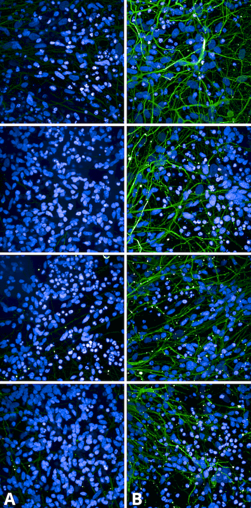
Immunocytochemistry/Immunofluorescence analysis using Rabbit Anti-Tau Monoclonal Antibody, Clone AH36 (SMC-601). Tissue: iPSC-derived cortical excitatory neurons. Species: Human. Primary Antibody: Rabbit Anti-Tau Monoclonal Antibody (SMC-601) at 1:500 for Overnight. Secondary Antibody: Donkey anti-rabbit: Alexa Fluor 488 at 1:1000. Counterstain: DAPI. A) iPSC-derived neurons from non-demented control (NDC). B) iPSC-derived neurons from subject with P301L MAPT mutation. Images acquired using an automated Opera Phoenix system. Each field of view is a max projection from 10 planes of 1 μm stacks.. Courtesy of: Francesco Paonessa.
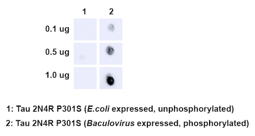
Dot Blot analysis using Rabbit Anti-Tau Monoclonal Antibody, Clone AH36 (SMC-601). Species: E. Coli, Baculovirus. Primary Antibody: Rabbit Anti-Tau Monoclonal Antibody (SMC-601) at 1:500. Secondary Antibody: Goat anti-rabbit IgG:HRP.
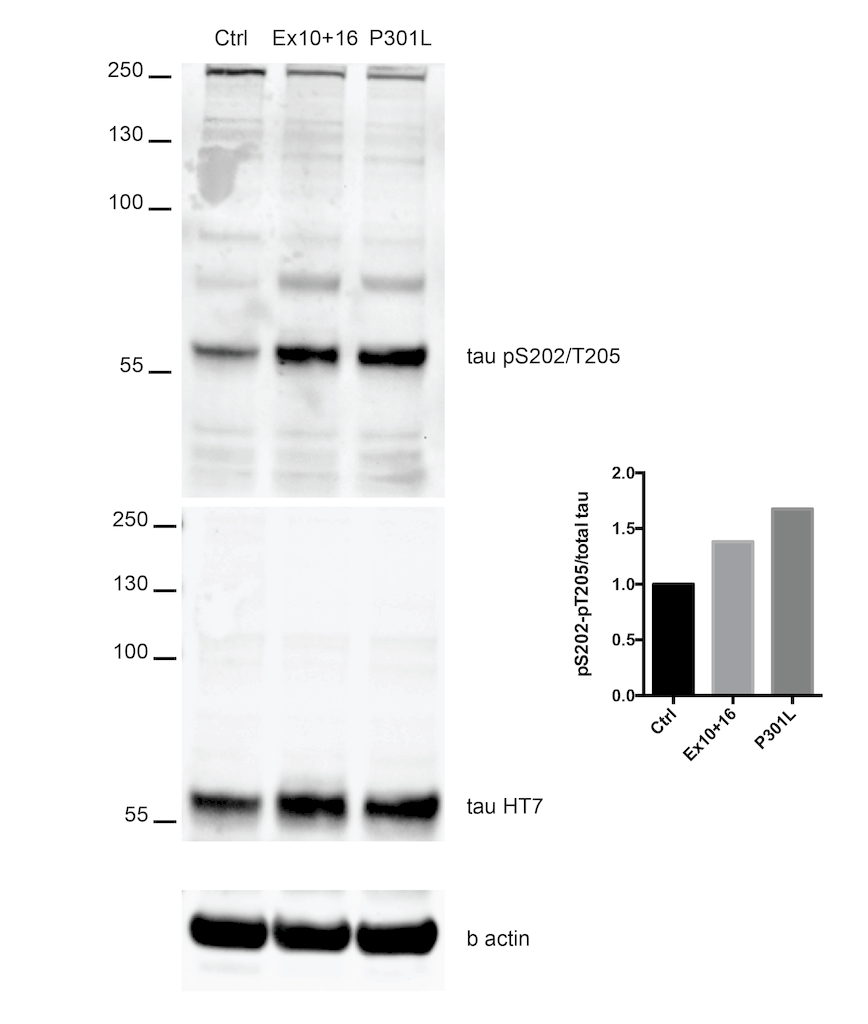
Western Blot analysis of Human iPSC-derived cortical neurons showing detection of Tau protein using Rabbit Anti-Tau Monoclonal Antibody, Clone AH36 (SMC-601). Lane 1: MW ladder. Lane 2: Control (non-disease) line. Lane 2: Ex10+16 tau mutant sample. Lane 3: P301L tau mutant sample. . Load: 50ug. Primary Antibody: Rabbit Anti-Tau Monoclonal Antibody (SMC-601) at 1:500 for Overnight. pSer202/pThr205 was detected using SMC-601. Total tau was detected using mouse anti-tau antibody (clone HT7). The bar graph on the right shows quantification of pSer202/pThr205 compared to total tau in each sample.. Courtesy of: Francesco Paonessa.
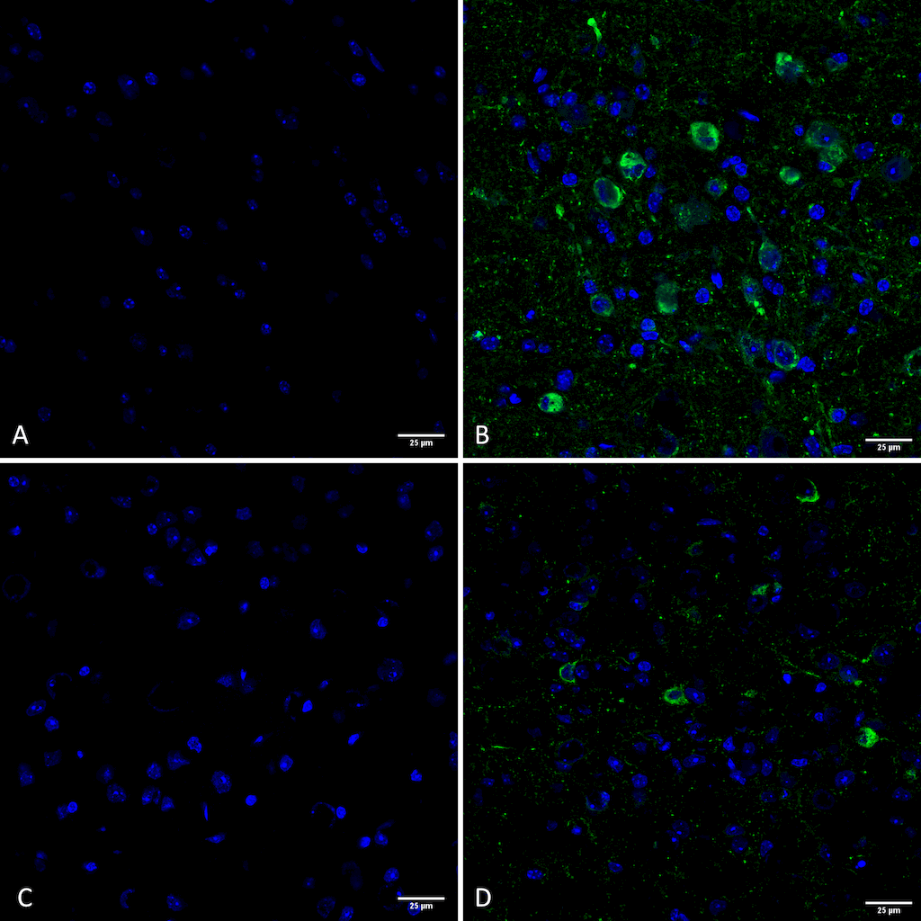
Immunohistochemistry analysis using Rabbit Anti-Tau Monoclonal Antibody, Clone AH36 (SMC-601). Tissue: Brain slice. Species: Mouse. Primary Antibody: Rabbit Anti-Tau Monoclonal Antibody (SMC-601) at 1:500 for Overnight at 4C. Secondary Antibody: Anti-Rabbit IgG: AlexaFluor 488. Counterstain: DAPI at 1:1000 for 5 min. (A) Pons of Non-Tg mouse. (B) Pons of P301SxUBQLN2 Tg mouse. (C) Prefrontal cortex of Non-Tg mouse. (D) Prefrontal cortex of P301SxUBQLN2 Tg mouse. IHC Protocol: 1. Post-fix brains in 4% PFA for 24 hours and put through a 10-30% sucrose gradient. 2. Section by cryostat at 10 uM thickness. 3. Fix in MeOH 15 min. 4. 3×10 min wash in PBS 1X. 5. Heat via microwave in 10mM Citrate Buffer, pH 6 for 4 min at power level 20. 6. Cool in solution for 20 min. 7. Wash 2×5 min in PBS. 8. Permeabilize in 0.5% Triton-X 100 in PBS 10 min. 9. Wash in PBS 10 min. 10. Block for 1 hour in 5% goat serum. 11. Incubate primary Ab (SMC-601 at 1:500) in blocking solution overnight at 4C. 12. Wash 3×10 min in PBS. 13. Incubate in secondary Ab Rb IgG Alexa-fluor 488. 14. Wash 3×10 min in PBS. 15. Incubate in DAPI 1:1000 for 5 min. 16. Wash 3×5 min. 17. Coverslip with Prolong-Gold.. Courtesy of: Julia Gerson, University of Michigan.
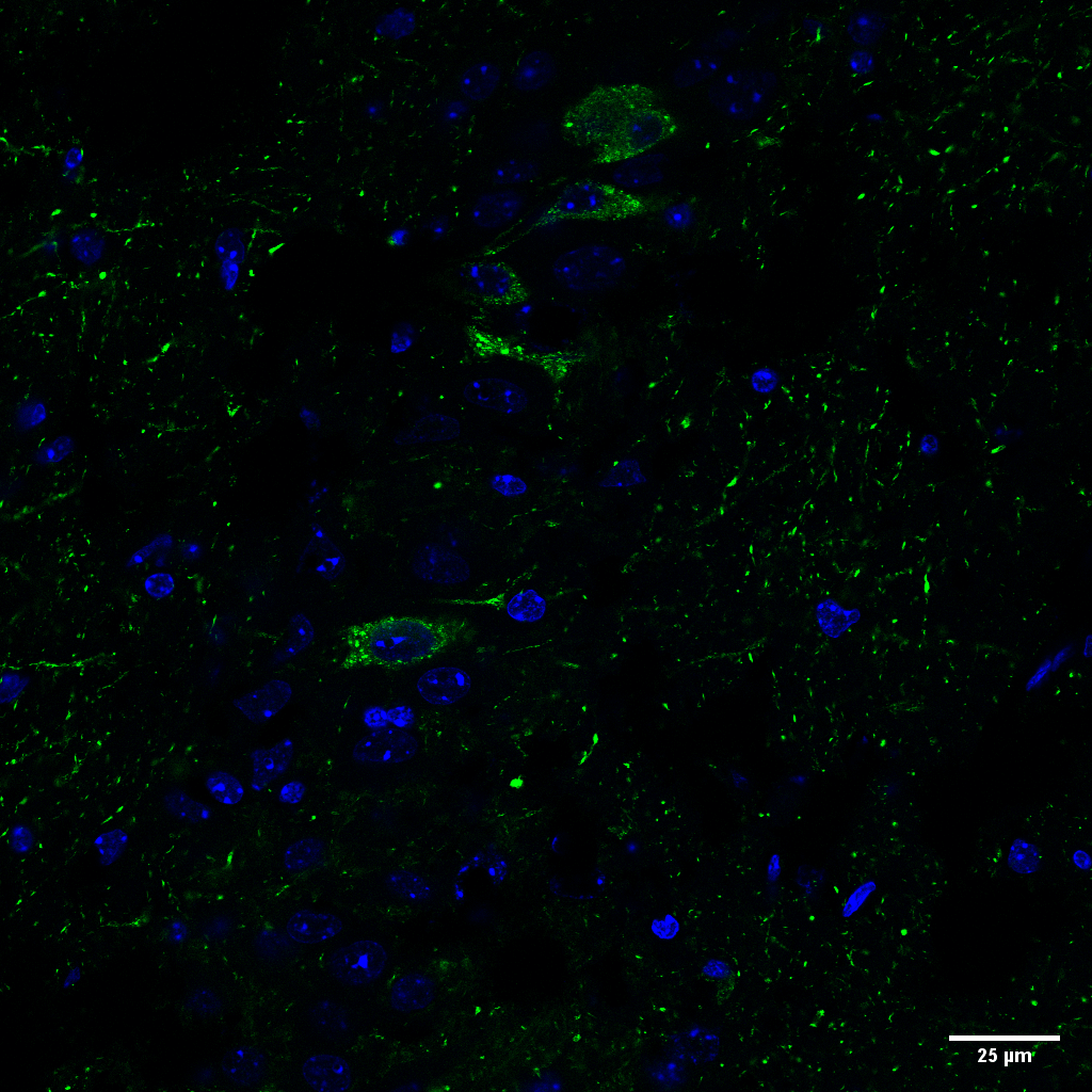
Immunohistochemistry analysis using Rabbit Anti-Tau Monoclonal Antibody, Clone AH36 (SMC-601). Tissue: Brain slice. Species: Mouse. Primary Antibody: Rabbit Anti-Tau Monoclonal Antibody (SMC-601) at 1:500 for Overnight at 4C. Secondary Antibody: Anti-Rabbit IgG: AlexaFluor 488. Counterstain: DAPI at 1:1000 for 5 min. CA3 Region of P301SxUBQLN2 Tg mouse. IHC Protocol: 1. Post-fix brains in 4% PFA for 24 hours and put through a 10-30% sucrose gradient. 2. Section by cryostat at 10 uM thickness. 3. Fix in MeOH 15 min. 4. 3×10 min wash in PBS 1X. 5. Heat via microwave in 10mM Citrate Buffer, pH 6 for 4 min at power level 20. 6. Cool in solution for 20 min. 7. Wash 2×5 min in PBS. 8. Permeabilize in 0.5% Triton-X 100 in PBS 10 min. 9. Wash in PBS 10 min. 10. Block for 1 hour in 5% goat serum. 11. Incubate primary Ab (SMC-601 at 1:500) in blocking solution overnight at 4C. 12. Wash 3×10 min in PBS. 13. Incubate in secondary Ab Rb IgG Alexa-fluor 488. 14. Wash 3×10 min in PBS. 15. Incubate in DAPI 1:1000 for 5 min. 16. Wash 3×5 min. 17. Coverslip with Prolong-Gold.. Courtesy of: Julia Gerson, University of Michigan.
 Powered by Bioz
StressMarq Biosciences :
Based on validation through cited publications.