
Advancing the Frontiers of Neurodegenerative Disease Research

Properties
| Storage Buffer | PBS pH7.4, 50% glycerol, 0.09% sodium azide *Storage buffer may change when conjugated |
| Storage Temperature | -20ºC, Conjugated antibodies should be stored according to the product label |
| Shipping Temperature | Blue Ice or 4ºC |
| Purification | Protein G Purified |
| Clonality | Monoclonal |
| Clone Number | N29/29 (Formerly sold as S29-29) |
| Isotype | IgG1 |
| Specificity | Detects ~60kDa. |
| Cite This Product | VGLUT2 Antibody (StressMarq Biosciences | Victoria, BC CANADA, Catalog# SMC-395, RRID: AB_11229706) |
| Certificate of Analysis | 1 µg/ml of SMC-395 was sufficient for detection of VGLut2 in 20 µg of rat brain lysate by colorimetric immunoblot analysis using goat anti-mouse IgG:HRP as the secondary antibody. |
Biological Description
| Alternative Names | VGLUT2, VGluT2, VGLU2, SLC17A6, Solute Carrier Family 17 Member 6, Solute Carrier Family 17 (Sodium-Dependent Inorganic Phosphate Cotransporter) Member 6, Solute Carrier Family 17 (Vesicular Glutamate Transporter) Member 6, Vesicular Glutamate Transporter 2, DNPI, Differentiation-Associated Na(+)-Dependent Inorganic Phosphate Cotransporter, Differentiation-Associated Na-Dependent Inorganic Phosphate Cotransporter, Differentiation-Associated BNPI, Sodium-Dependent Inorganic Phosphate Cotransporter |
| Research Areas | Cell Markers, Cell Signaling, Neuron Markers, Neuroscience, Neurotransmitter Transporters, Presynaptic Markers, Pumps/Transporters |
| Cellular Localization | Cell Junction, Cytoplasmic Vesicle, Membrane, Secretory vesicle, Synapse, Synaptic vesicle membrane |
| Accession Number | NP_445879.1 |
| Gene ID | 84487 |
| Swiss Prot | Q9JI12 |
| Scientific Background |
Vesicular Glutamate Transporter 2 (VGLUT2), encoded by SLC17A6, is an ATP-dependent, chloride-sensitive transporter essential for loading glutamate into synaptic vesicles. Alongside VGLUT1 (BNPI) and VGLUT3, VGLUT2 plays a pivotal role in excitatory neurotransmission. Its expression is regionally distinct and complementary to VGLUT1: while VGLUT1 predominates in telencephalic regions, VGLUT2 is highly expressed in the diencephalon and lower brainstem—areas critical for autonomic control, sensory integration, and motor coordination. VGLUT2 is also expressed in rat pinealocytes and pancreatic α and β cells, where it responds to glucose levels, suggesting a broader role in neuroendocrine signaling. This transporter is particularly relevant in the context of neurodegenerative diseases, as its dysregulation has been implicated in conditions such as Parkinson’s disease, amyotrophic lateral sclerosis (ALS), and Huntington’s disease. Its involvement in glutamate homeostasis and excitotoxicity positions VGLUT2 as a key molecular target for understanding disease mechanisms and developing therapeutic interventions. Although VGLUT3 shares approximately 72% sequence homology with VGLUT2 and VGLUT1, each isoform exhibits distinct spatial and functional profiles. VGLUT2’s unique distribution and regulatory mechanisms make it a critical focus in neuroscience research, particularly in studies of synaptic plasticity, circuit dysfunction, and neurodegeneration. |
| References |
1. Aihara Y., et al. (2000) J Neurochem. 74: 2622-2625. 2. Kaneko T., and Fujiyama F. (2002) Neurosci Res. 42: 243-250. 3. Gras C., et al. (2002) J Neurosci. 22: 5442-5451. 4. Takamori S., Malherbe P., Broger C., and Jahn R. (2002) EMBO Rep. 3: 798-803. 5. Morimoto R., Hayashi M., Yatsushiro S., Otsuka M., Yamamoto A., and Moriyama Y. (2003) J Neurochem. 84: 382-391. 6. Bai L., Zhang X., and Ghishan F.K. (2003) Am. J. Physiol. Gastrointest. Liver Physiol. 284: G808-814. |
Product Images
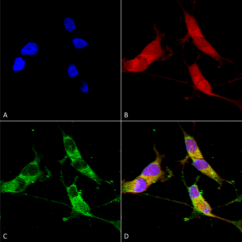
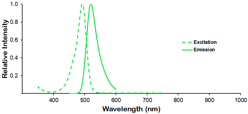
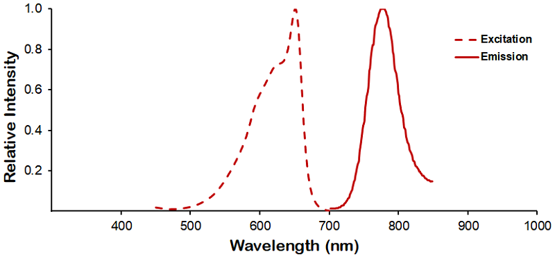
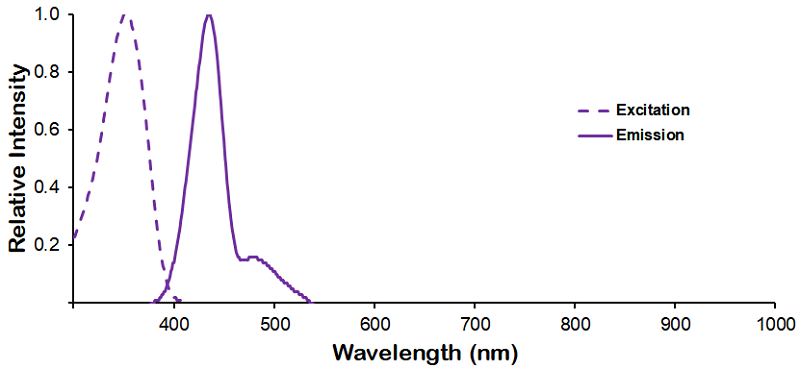
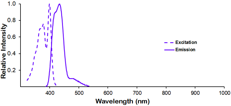
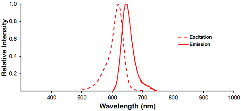
Immunocytochemistry/Immunofluorescence analysis using Mouse Anti-VGLUT2 Monoclonal Antibody, Clone N29/29 (SMC-395). Tissue: Neuroblastoma cells (SH-SY5Y). Species: Human. Fixation: 4% PFA for 15 min. Primary Antibody: Mouse Anti-VGLUT2 Monoclonal Antibody (SMC-395) at 1:50 for overnight at 4°C with slow rocking. Secondary Antibody: AlexaFluor 488 at 1:1000 for 1 hour at RT. Counterstain: Phalloidin-iFluor 647 (red) F-Actin stain; Hoechst (blue) nuclear stain at 1:800, 1.6mM for 20 min at RT. (A) Hoechst (blue) nuclear stain. (B) Phalloidin-iFluor 647 (red) F-Actin stain. (C) VGLUT2 Antibody (D) Composite.
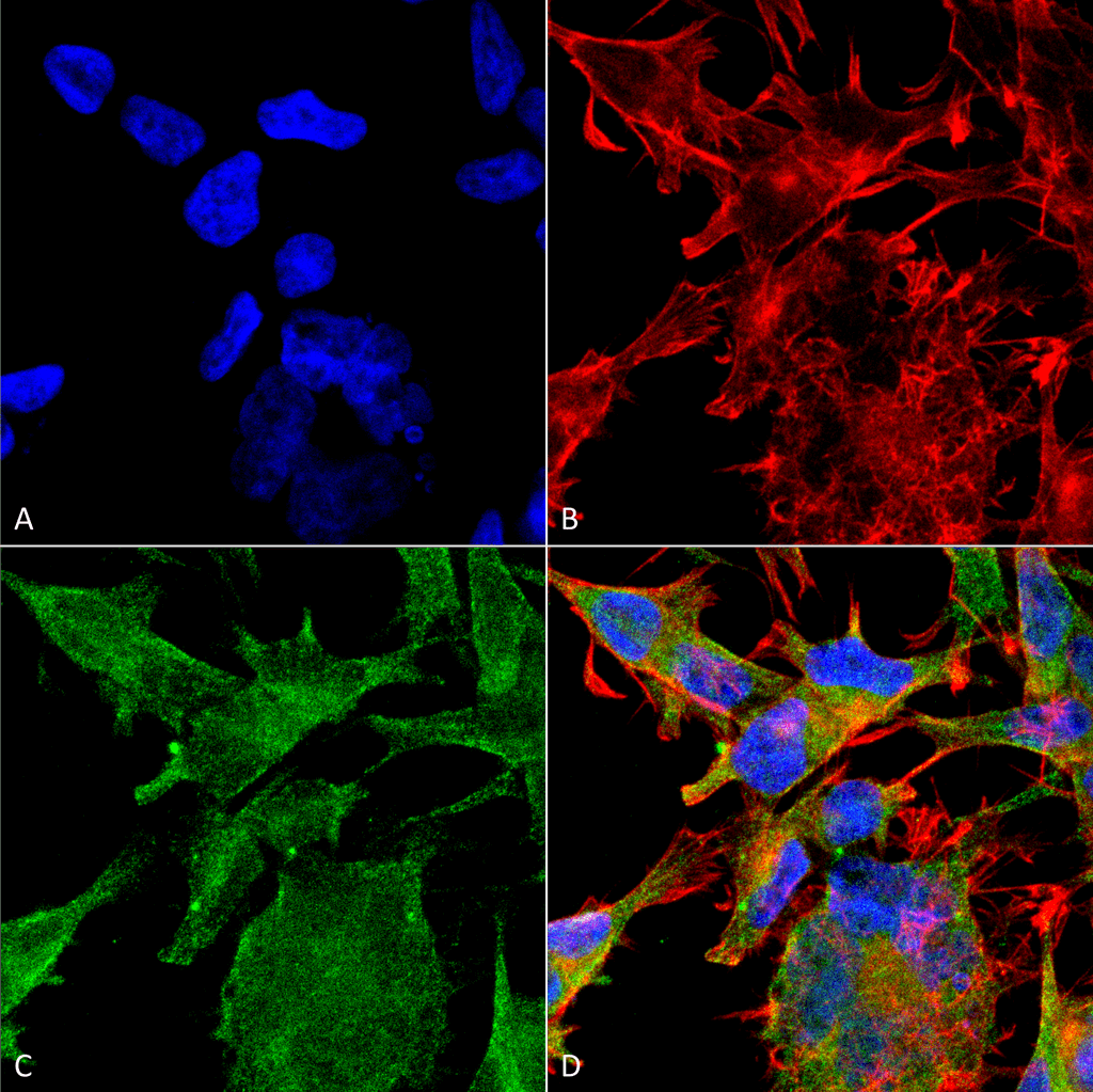
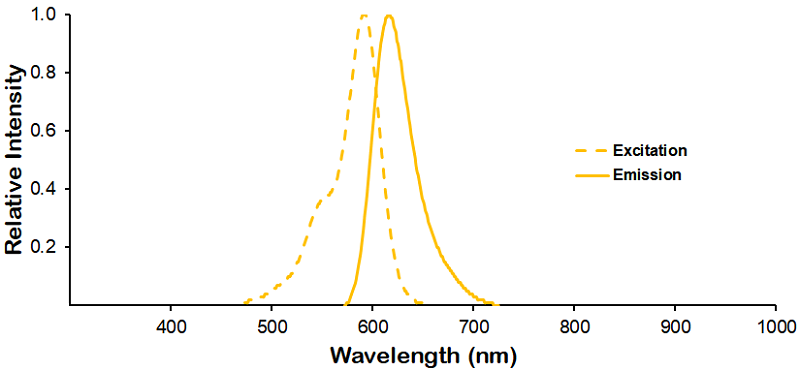
Immunocytochemistry/Immunofluorescence analysis using Mouse Anti-VGLUT2 Monoclonal Antibody, Clone N29/29 (SMC-395). Tissue: Neuroblastoma cell line (SK-N-BE). Species: Human. Fixation: 4% Formaldehyde for 15 min at RT. Primary Antibody: Mouse Anti-VGLUT2 Monoclonal Antibody (SMC-395) at 1:100 for 60 min at RT. Secondary Antibody: Goat Anti-Mouse ATTO 488 at 1:200 for 60 min at RT. Counterstain: Phalloidin Texas Red F-Actin stain; DAPI (blue) nuclear stain at 1:1000, 1:5000 for 60 min at RT, 5 min at RT. Localization: Cytoplasmic Vesicle, Secretory Vesicle, Synaptic Vesicle Membrane, Membrane, Cell Junction, Synapse. Magnification: 60X. (A) DAPI (blue) nuclear stain. (B) Phalloidin Texas Red F-Actin stain. (C) VGLUT2 Antibody. (D) Composite.
![<p>Western blot analysis with StressMarq’s Mouse Anti-Rat VGlut2 monoclonal antibody, clone [N29/29] (SMC-395) showing detection of VGlut2 protein in Rat Brain Membrane at ~60 kDa. Block: 5% skim milk for 1 hr at RT. Primary Antibody: Mouse Anti-VGlut2 Monoclonal Antibody (SMC-395) at 1:1000 for 2 hrs at RT. Secondary Antibody: Goat anti-mouse IgG:HRP at 1:5000 for 1 hr at RT. Color Development: Chemiluminescent for HRP (Moss) for 1 min at RT. Exposed 15 seconds.</p>](https://www.stressmarq.com/wp-content/uploads/SMC-395_VGLUT2_Antibody_N29-29_WB_Rat_brain-membrane-lysate_1.png)
Western blot analysis with StressMarq’s Mouse Anti-Rat VGlut2 monoclonal antibody, clone [N29/29] (SMC-395) showing detection of VGlut2 protein in Rat Brain Membrane at ~60 kDa. Block: 5% skim milk for 1 hr at RT. Primary Antibody: Mouse Anti-VGlut2 Monoclonal Antibody (SMC-395) at 1:1000 for 2 hrs at RT. Secondary Antibody: Goat anti-mouse IgG:HRP at 1:5000 for 1 hr at RT. Color Development: Chemiluminescent for HRP (Moss) for 1 min at RT. Exposed 15 seconds.
 Powered by Bioz
Reviews
There are no reviews yet.