Discovery through Partnership | Excellence through Quality


Properties
| Storage Buffer | PBS pH7.2, 50% glycerol, 0.09% sodium azide *Storage buffer may change when conjugated |
| Storage Temperature | -20ºC, Conjugated antibodies should be stored according to the product label |
| Shipping Temperature | Blue Ice or 4ºC |
| Purification | Peptide Affinity Purified |
| Clonality | Polyclonal |
| Specificity | Detects the C-terminal domain of Calnexin ~90kDa. Weak detection in Chicken, Drosophila, and Xenopus tissues. |
| Cite This Product | StressMarq Biosciences Cat# SPC-182, RRID: AB_2068990 |
| Certificate of Analysis | A 1:2000 dilution of SPC-182 was sufficient for detection of Calnexin in 10 µg of HeLa cell lysate by ECL immunoblot analysis. |
Biological Description
| Alternative Names | Calnexin, CALX_HUMAN, CANX, CNX, FLJ26570, IP90, p88, p90, Histocompatibility complex class I antigen binding protein p88, Major histocompatibility complex class I antigen-binding protein p88 |
| Research Areas | Cell Signaling, Organelle Markers, Tags and Cell Markers |
| Cellular Localization | Endoplasmic Reticulum, Endoplasmic reticulum membrane, Melanosome |
| Accession Number | NP_001003232.1 |
| Gene ID | 403908 |
| Swiss Prot | P24643 |
| Scientific Background |
Calnexin is a ~90 kDa integral membrane protein of the endoplasmic reticulum (ER), also known as IP90, p88, or p90. Structurally, it comprises a large N-terminal calcium-binding luminal domain (~50 kDa), a single transmembrane helix, and a short, acidic cytoplasmic tail (CT). Unlike many ER-resident proteins that rely on a C-terminal KDEL motif for retention, Calnexin is retained in the ER via positively charged residues in its cytosolic tail. Functionally, Calnexin is a central component of the ER quality control system. It acts as a molecular chaperone, facilitating the proper folding and assembly of nascent glycoproteins. In concert with calreticulin, Calnexin binds to monoglucosylated N-linked glycans on folding intermediates, ensuring fidelity in glycoprotein maturation. It also interacts with key immune molecules, including MHC class I heavy chains, T cell receptor subunits, and B cell immunoglobulins. In neuroscience, Calnexin-CT is gaining attention for its role in neurodegenerative disease. ER stress and impaired protein folding are hallmarks of disorders such as Alzheimer’s, Parkinson’s, and ALS. Dysregulation of Calnexin function can exacerbate proteostasis imbalance, leading to accumulation of misfolded proteins and neuronal dysfunction. Given its dual role in protein folding and ER homeostasis, Calnexin—particularly its cytoplasmic tail—represents a promising target for therapeutic strategies aimed at restoring proteostasis in neurodegenerative diseases. |
| References |
1. Rajagopalan S., Xu Y., and Brenner M.B. (1994) Science. 263(5145): 387-90. 2. Tjoelker L.W., et al. (1994) Biochemistry. 33: 3229. 3. Schrag J. et al. (2001) Molecular Cell. 8(3): 633-644. 4. Janiszewski M. (2005) J. Biol Chem. 280(49): 40813-40819. 5. Elagoz A., Callejo M., Armstrong J., and Rokeach L. A. (1999) J. Cell Sci. 112: 4449-4460. 6. Otteken A. and Moss B. (1996) J Bio Chem. 271(1): 97-103. 7. Galvin K. et al. (1992) Proc Natl Acad Sci USA. 89(18): 8452-6. |
Product Images
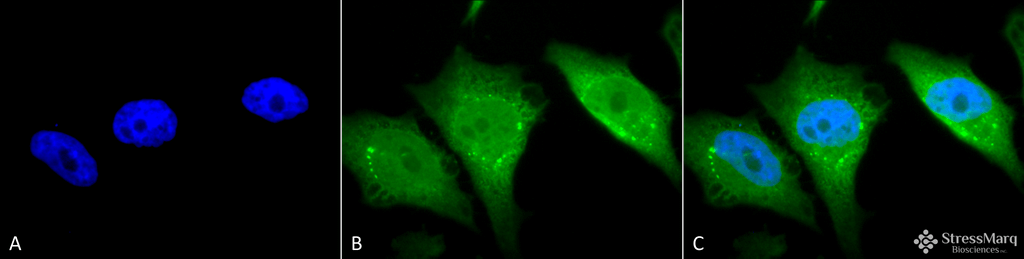
Immunocytochemistry/Immunofluorescence analysis using Rabbit Anti-Calnexin-CT Polyclonal Antibody (SPC-182). Tissue: Heat Shocked Cervical cancer cell line (HeLa). Species: Human. Fixation: 2% Formaldehyde for 20 min at RT. Primary Antibody: Rabbit Anti-Calnexin-CT Polyclonal Antibody (SPC-182) at 1:80 for 12 hours at 4°C. Secondary Antibody: FITC Goat Anti-Rabbit (green) at 1:200 for 2 hours at RT. Counterstain: DAPI (blue) nuclear stain at 1:40000 for 2 hours at RT. Localization: Endoplasmic reticulum membrane. Melanosome. Magnification: 100x. Heat Shocked at 42°C for 1h.
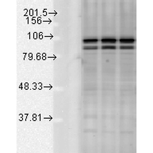
Western blot analysis of Rat tissue mix showing detection of Calnexin-CT protein using Rabbit Anti-Calnexin-CT Polyclonal Antibody (SPC-182). Load: 15 µgprotein. Block: 1.5% BSA for 30 minutes at RT. Primary Antibody: Rabbit Anti-Calnexin-CT Polyclonal Antibody (SPC-182) at 1:1000 for 2 hours at RT. Secondary Antibody: Donkey Anti-Rabbit IgG: HRP for 1 hour at RT.
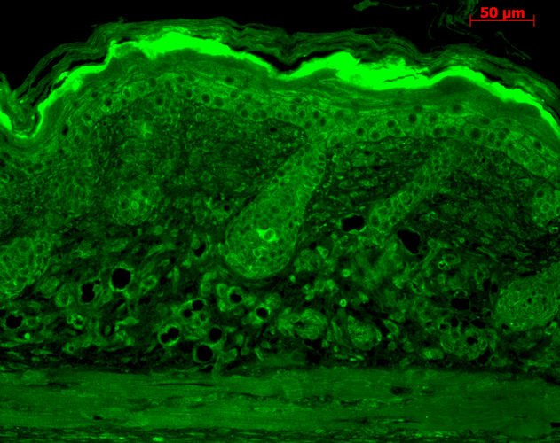
Immunohistochemistry analysis using Rabbit Anti-Calnexin-CT Polyclonal Antibody (SPC-182). Tissue: backskin. Species: Mouse. Fixation: Bouin’s Fixative Solution. Primary Antibody: Rabbit Anti-Calnexin-CT Polyclonal Antibody (SPC-182) at 1:100 for 1 hour at RT. Secondary Antibody: FITC Goat Anti-Rabbit (green) at 1:50 for 1 hour at RT. Localization: Hair Follicles, Basal cells in epidermis, and second layer of epidermis.
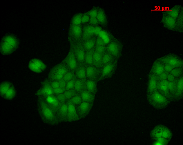
Immunocytochemistry/Immunofluorescence analysis using Rabbit Anti-Calnexin-CT Polyclonal Antibody (SPC-182). Tissue: HaCaT cells. Species: Human. Fixation: Cold 100% methanol at -20C for 10 minutes. Primary Antibody: Rabbit Anti-Calnexin-CT Polyclonal Antibody (SPC-182) at 1:100 for 12 hours at 4°C. Secondary Antibody: FITC Goat Anti-Rabbit at 1:50 for 1-2 hours at RT in dark. Localization: Nuclear staining, cytoplasmic staining.
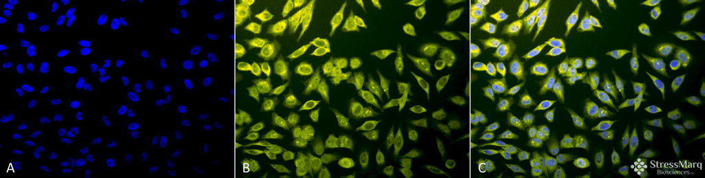
Immunocytochemistry/Immunofluorescence analysis using Rabbit Anti-Calnexin-CT Polyclonal Antibody (SPC-182). Tissue: Heat Shocked Cervical cancer cell line (HeLa). Species: Human. Fixation: 2% Formaldehyde for 20 min at RT. Primary Antibody: Rabbit Anti-Calnexin-CT Polyclonal Antibody (SPC-182) at 1:80 for 12 hours at 4°C. Secondary Antibody: R-PE Goat Anti-Rabbit (yellow) at 1:200 for 2 hours at RT. Counterstain: DAPI (blue) nuclear stain at 1:40000 for 2 hours at RT. Localization: Endoplasmic reticulum membrane. Melanosome. Magnification: 20x. (A) DAPI (blue) nuclear stain. (B) Anti-Calnexin-CT Antibody. (C) Composite. Heat Shocked at 42°C for 1h.
 Powered by Bioz
Reviews
There are no reviews yet.