Discovery through Partnership | Excellence through Quality


Properties
| Storage Buffer | PBS pH7.4, 50% glycerol, 0.09% sodium azide *Storage buffer may change when conjugated |
| Storage Temperature | -20ºC, Conjugated antibodies should be stored according to the product label |
| Shipping Temperature | Blue Ice or 4ºC |
| Purification | Peptide Affinity Purified |
| Clonality | Polyclonal |
| Specificity | Detects ~44kda (ERK1) and ~42kDa (ERK2). |
| Cite This Product | StressMarq Biosciences Cat# SPC-120, RRID: AB_2266080 |
| Certificate of Analysis | A 1:1000 dilution of SPC-120 was sufficient for detection of ERK1/2 in 20 µg of HeLa cell lysate by ECL immunoblot analysis. |
Biological Description
| Alternative Names | ERK1, ERK2, ERT1, ERT2, MAP kinase 1, MAP kinase 2, MAPK1, MAPK2, MAPK3, p38, p40, p41, p41mapk, p42 MAPK, p44 ERK1, p44 MAPK, PRKM1, PRKM2, PRKM3 |
| Research Areas | Alzheimer's Disease, Cell Signaling, Neurodegeneration, Neuroscience, Phosphorylation, Post-translational Modifications |
| Cellular Localization | Cytoplasm, Nucleus |
| Accession Number | NP_059043.1 |
| Gene ID | 50689 |
| Swiss Prot | P21708 |
| Scientific Background |
Extracellular signal-regulated kinases 1 and 2 (Erk1/2) are central components of the MAPK signaling cascade, orchestrating cellular responses to growth factors, stress, and synaptic activity. In the nervous system, Erk1/2 regulate neuronal differentiation, synaptic plasticity, and memory formation. Dysregulation of Erk1/2 signaling has been implicated in the pathogenesis of multiple neurodegenerative diseases, including Alzheimer’s, Parkinson’s, and Huntington’s disease. Aberrant Erk1/2 activation can lead to chronic neuroinflammation, oxidative stress, and tau hyperphosphorylation—hallmarks of neurodegeneration. Conversely, controlled Erk1/2 signaling supports neuroprotection, axonal regeneration, and cognitive resilience. This dual role positions Erk1/2 as both a biomarker and a therapeutic target in neurodegenerative research. As precision medicine advances, modulating Erk1/2 activity offers a promising strategy to restore neuronal function and delay disease progression. |
| References |
1. Boulton TG. et al. (1991) Biochemistry. 30(1):278-86. 2. Yoon S., and Seger R. (2006) Growth Factors 24:21-44. 3. Wolf G. (2005) Antioxid Redox Signal 7:1337-1345. 4. Chuerland D., Marmor G., Shainskaya A. and Seger R. (2008) J Biol Chem. Epub: http://www.jbc.org/cgi/doi/10.1074/jbc.M709030200 5. Zhuang S., and Schnellmann R.G. (2006) J Pharmacol Exp Ther 319:991-997. |
Product Images
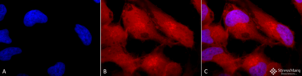
Immunocytochemistry/Immunofluorescence analysis using Rabbit Anti-Erk1/2 Polyclonal Antibody (SPC-120). Tissue: Cervical cancer cell line (HeLa). Species: Human. Fixation: 2% Formaldehyde for 20 min at RT. Primary Antibody: Rabbit Anti-Erk1/2 Polyclonal Antibody (SPC-120) at 1:100 for 12 hours at 4°C. Secondary Antibody: APC Goat Anti-Rabbit (red) at 1:200 for 2 hours at RT. Counterstain: DAPI (blue) nuclear stain at 1:40000 for 2 hours at RT. Localization: Cytoplasm. Nucleus. Magnification: 100x. (A) DAPI (blue) nuclear stain. (B) Anti-Erk1/2 Antibody. (C) Composite.
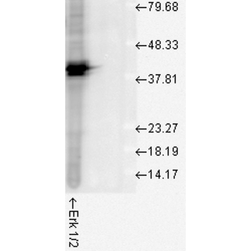
Western blot analysis of Human Cell line lysates showing detection of ERK1 protein using Rabbit Anti-ERK1 Polyclonal Antibody (SPC-120). Load: 15 µgprotein. Block: 1.5% BSA for 30 minutes at RT. Primary Antibody: Rabbit Anti-ERK1 Polyclonal Antibody (SPC-120) at 1:1000 for 2 hours at RT. Secondary Antibody: Donkey Anti-Rabbit IgG: HRP for 1 hour at RT.
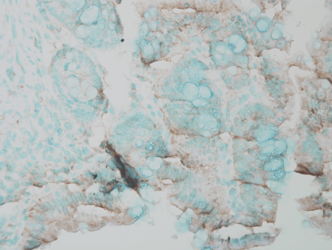
Immunohistochemistry analysis using Rabbit Anti-ERK1 Polyclonal Antibody (SPC-120). Tissue: Inflamed colon. Species: Mouse. Fixation: Formalin. Primary Antibody: Rabbit Anti-ERK1 Polyclonal Antibody (SPC-120) at 1:25000 for 12 hours at 4°C. Secondary Antibody: Biotin Goat Anti-Rabbit at 1:2000 for 1 hour at RT. Counterstain: Methyl Green at 200uL for 2 min at RT. This image was produced using an amplifying IHC wash buffer. The antibody has therefore been diluted more than is recommended for other applications..
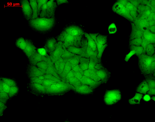
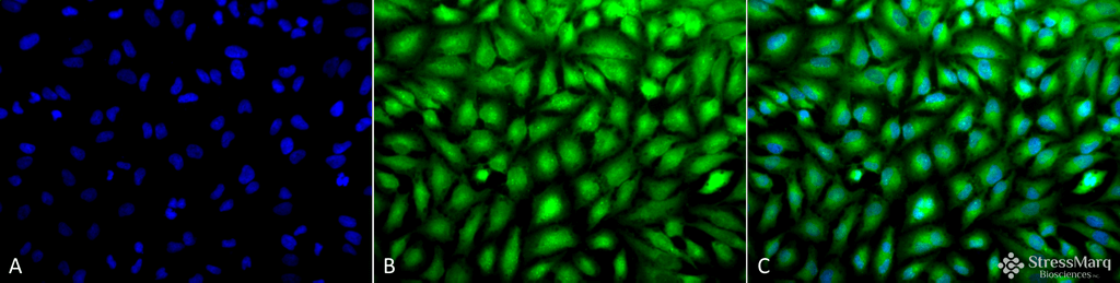
Immunocytochemistry/Immunofluorescence analysis using Rabbit Anti-ERK1 Polyclonal Antibody (SPC-120). Tissue: HaCaT cells. Species: Human. Fixation: Cold 100% methanol at -20C for 10 minutes. Primary Antibody: Rabbit Anti-ERK1 Polyclonal Antibody (SPC-120) at 1:100 for 12 hours at 4°C. Secondary Antibody: FITC Goat Anti-Rabbit at 1:50 for 1-2 hours at RT in dark. Localization: Cytoplasm. Nucleus.
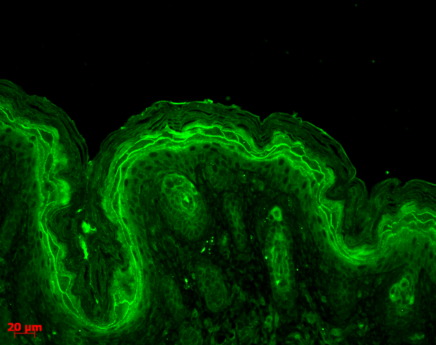
Immunohistochemistry analysis using Rabbit Anti-ERK1 Polyclonal Antibody (SPC-120). Tissue: backskin. Species: Mouse. Fixation: Bouin’s Fixative Solution. Primary Antibody: Rabbit Anti-ERK1 Polyclonal Antibody (SPC-120) at 1:100 for 1 hour at RT. Secondary Antibody: FITC Goat Anti-Rabbit (green) at 1:50 for 1 hour at RT. Localization: Cytoplasm.
Immunocytochemistry/Immunofluorescence analysis using Rabbit Anti-Erk1/2 Polyclonal Antibody (SPC-120). Tissue: Cervical cancer cell line (HeLa). Species: Human. Fixation: 2% Formaldehyde for 20 min at RT. Primary Antibody: Rabbit Anti-Erk1/2 Polyclonal Antibody (SPC-120) at 1:100 for 12 hours at 4°C. Secondary Antibody: FITC Goat Anti-Rabbit (green) at 1:200 for 2 hours at RT. Counterstain: DAPI (blue) nuclear stain at 1:40000 for 2 hours at RT. Localization: Cytoplasm. Nucleus. Magnification: 20x. (A) DAPI (blue) nuclear stain. (B) Anti-Erk1/2 Antibody. (C) Composite.
 Powered by Bioz
Comparison Analysis from AntibodyResource.com :
Anti-ERK1/2 polyclonal antibody (Catalog No. SPC-120, referenced as P132 in the report) was independently validated for use in western blot on human breast adenocarcinoma (MCF7 cell line) whole cell lysate and mouse embryonic fibroblast (NIH3T3 cell line) whole cell lysate at a dilution of 1:5000, with bands detected at the expected molecular weight of 38-43 kDa.Read the full ERK1 antibody comparison report (PDF) prepared by AntibodyResource.comAntibody Resource is spearheading an initiative designed to compare antibodies from numerous suppliers using identical samples/tissues and an identical protocol. In doing so, they hope to enable scientists to form an unrivalled opinion of which is the most suitable antibody for their research and which is going to require the least amount of optimization, a process which can often take weeks or months. For the purposes of the antibody comparison initiative, the best antibodies from each manufacturer were selected and compared side-by-side using the same experimental conditions to provide a direct comparison. The antibodies were collected centrally, repackaged and given an internal reference ID prior to delivery to independent laboratories to ensure objective testing and to minimize bias.“
StressMarq Biosciences :
Based on validation through cited publications.