Discovery through Partnership | Excellence through Quality


Properties
| Storage Buffer | PBS pH7.4, 50% glycerol, 0.09% sodium azide *Storage buffer may change when conjugated |
| Storage Temperature | -20ºC, Conjugated antibodies should be stored according to the product label |
| Shipping Temperature | Blue Ice or 4ºC |
| Purification | Peptide Affinity Purified |
| Clonality | Polyclonal |
| Specificity | Detects ~78kDa. |
| Cite This Product | StressMarq Biosciences Cat# SPC-107, RRID: AB_2119986 |
| Certificate of Analysis | A 1:1000 dilution of SPC-107 was sufficient for detection of Grp78 in 10 µg of rat tissue lysate by ECL immunoblot analysis. |
Biological Description
| Alternative Names | GRP78, GRP-78, Grp78, GRP 78, HSPA5, HSPA 5, Heat shock 70 kDa protein 5, Heat Shock 70kDa Protein 5, BiP, BIP, Binding immunoglobulin protein, Immunoglobulin heavy chain-binding protein, Immunoglobulin Heavy Chain Binding Protein, Endoplasmic reticulum lumenal Ca(2+)-binding protein grp78, Endoplasmic reticulum lumenal, Ca2+ binding protein grp78, Glucose Regulated Protein 78kDa, 78 kDa glucose-regulated protein, 78 kDa glucose regulated protein, HSCe70, MIF2, mBiP, Sez7, AL022860, AU019543, D2Wsu141e, D2Wsu17e, FLJ26106, GRP78_HUMAN |
| Research Areas | Cancer, Cell Signaling, Chaperone Proteins, Heat Shock, Organelle Markers, Protein Trafficking, Tags and Cell Markers |
| Cellular Localization | Cytoplasm, Endoplasmic Reticulum, Endoplasmic reticulum lumen, Melanosome |
| Accession Number | NP_037215.1 |
| Gene ID | 25617 |
| Swiss Prot | P06761 |
| Scientific Background |
GRP78 (also known as BiP) is a 78-kDa glucose-regulated protein and a master regulator of the unfolded protein response (UPR). As a key ER chaperone, it ensures proper protein folding, assembly, and the clearance of misfolded proteins, especially under stress conditions. Its expression is controlled by an internal ribosome entry site (IRES), which becomes highly active during cellular stress, such as heat shock or oxidative damage. GRP78 is essential for maintaining neuronal homeostasis and preventing apoptosis, making it a critical player in neurodegenerative disease contexts. In neuroscience, GRP78 has demonstrated neuroprotective effects against glutamate toxicity and oxidative stress. Its levels are notably reduced in the brains of Alzheimer’s disease patients, highlighting its potential as a biomarker and therapeutic target. Additionally, GRP78 supports embryonic development and pluripotent cell survival, further underscoring its importance in neural cell viability. However, its overexpression under extreme stress may contribute to drug resistance in neuro-oncology, adding complexity to its therapeutic modulation. |
| References |
1. Cho, S. et al. (2007). Mol Cell Biol 27(1): 368-83. 2. Yang, Y. et al. (1998) J Biol Chem 273: 25552-25555. 3. Luo, S. et al (2006) 26 (15): 5688-97. 4. Yu, Z. et al. (1999) Exp Neurol. 15: 302-314. 5. Koomagi, R. et al. (1999) Anticancer Res. 19:4333-4336. 6. Laquerre, S. et al. (1998) J. Virology 72: 4940-4949. 7. Dong, D. et al. (2005) Cancer Res 65(13): 5785-91. |
Product Images
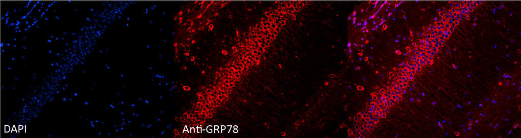
Immunocytochemistry/Immunofluorescence analysis using Rabbit Anti-GRP78 Polyclonal Antibody (SPC-107). Tissue: Hippocampal Section. Species: Mouse. Fixation: 4% Formaldehyde for 12 hours at RT. Paraffin embedded.. Primary Antibody: Rabbit Anti-GRP78 Polyclonal Antibody (SPC-107) at 1:100 for 12 hours at 4°C. Secondary Antibody: Alexa Fluor 555 Goat Anti-Rabbit at 1:250 for 1 hour at RT. Counterstain: Hoechst at 1:1000 for 10 min at RT. Localization: Grp78 staining in mouse pyramidal cell layer.. Magnification: 20x. Slice thickness: 7 microns. Courtesy of: Rachel Reith, NIH/NIMH..
Western blot analysis of Human, Dog, Mouse Cell line lysates showing detection of GRP78 protein using Rabbit Anti-GRP78 Polyclonal Antibody (SPC-107). Primary Antibody: Rabbit Anti-GRP78 Polyclonal Antibody (SPC-107) at 1:1000.
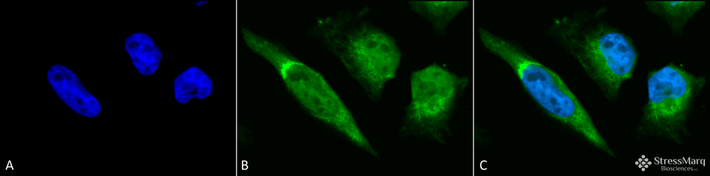
Immunocytochemistry/Immunofluorescence analysis using Rabbit Anti-GRP78 Polyclonal Antibody (SPC-107). Tissue: Heat Shocked Cervical cancer cell line (HeLa). Species: Human. Fixation: 2% Formaldehyde for 20 min at RT. Primary Antibody: Rabbit Anti-GRP78 Polyclonal Antibody (SPC-107) at 1:100 for 12 hours at 4°C. Secondary Antibody: FITC Goat Anti-Rabbit (green) at 1:200 for 2 hours at RT. Counterstain: DAPI (blue) nuclear stain at 1:40000 for 2 hours at RT. Localization: Endoplasmic reticulum lumen. Melanosome. Cytoplasm . Magnification: 100x. (A) DAPI (blue) nuclear stain. (B) Anti-GRP78 Antibody. (C) Composite. Heat Shocked at 42°C for 30 min.
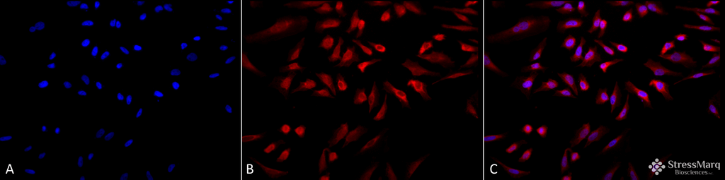
Immunocytochemistry/Immunofluorescence analysis using Rabbit Anti-GRP78 Polyclonal Antibody (SPC-107). Tissue: Heat Shocked Cervical cancer cell line (HeLa). Species: Human. Fixation: 2% Formaldehyde for 20 min at RT. Primary Antibody: Rabbit Anti-GRP78 Polyclonal Antibody (SPC-107) at 1:100 for 12 hours at 4°C. Secondary Antibody: APC Goat Anti-Rabbit (red) at 1:200 for 2 hours at RT. Counterstain: DAPI (blue) nuclear stain at 1:40000 for 2 hours at RT. Localization: Endoplasmic reticulum lumen. Melanosome. Cytoplasm . Magnification: 20x. (A) DAPI (blue) nuclear stain. (B) Anti-GRP78 Antibody. (C) Composite. Heat Shocked at 42°C for 30 min.
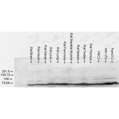
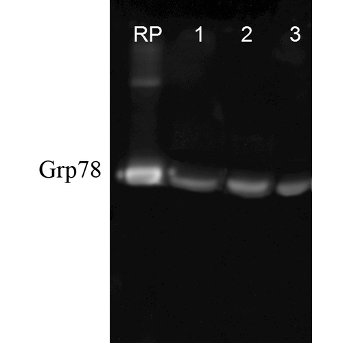
Western blot analysis of Rat Tissue lysates showing detection of GRP78 protein using Rabbit Anti-GRP78 Polyclonal Antibody (SPC-107). Load: 15 µgprotein. Block: 1.5% BSA. Primary Antibody: Rabbit Anti-GRP78 Polyclonal Antibody (SPC-107) at 1:1000 for 2 hours at RT. Secondary Antibody: Donkey Anti-Rabbit IgG: HRP for 1 hour at RT.
 Powered by Bioz
StressMarq Biosciences :
Based on validation through cited publications.