Discovery through Partnership | Excellence through Quality


Properties
| Storage Buffer | PBS pH7.4, 50% glycerol, 0.09% sodium azide *Storage buffer may change when conjugated |
| Storage Temperature | -20ºC, Conjugated antibodies should be stored according to the product label |
| Shipping Temperature | Blue Ice or 4ºC |
| Purification | Peptide Affinity Purified |
| Clonality | Polyclonal |
| Specificity | Detects ~10kDa. It also recognizes ubiquinated proteins. |
| Cite This Product | Ubiquitin Antibody (StressMarq Biosciences | Victoria, BC CANADA, Catalog# SPC-119, RRID: AB_2285248) |
| Certificate of Analysis | A 1:1000 dilution of SPC-119 was sufficient for detection of free ubiquitin in 15 µg of HeLa lysate by ECL immunoblot analysis using Donkey anti-rabbit IgG:HRP as the secondary antibody. |
Biological Description
| Alternative Names | Polyubiquitin B, RPS27A, UBA52, UBB, UBC, ubiquitin B |
| Research Areas | Alzheimer's Disease, Cell Signaling, Neurodegeneration, Neuroscience, Post-translational Modifications, Ubiquitination |
| Cellular Localization | Cell membrane, Cytoplasm, Nucleus |
| Accession Number | NP_776558.1 |
| Gene ID | 281370 |
| Swiss Prot | P0CG53 |
| Scientific Background |
Ubiquitin is a highly conserved, small regulatory protein that plays a pivotal role in maintaining cellular protein homeostasis through the ubiquitin-proteasome system (UPS) and autophagy pathways. By tagging damaged, misfolded, or excess proteins for degradation, ubiquitin ensures proper protein turnover and prevents toxic protein accumulation—an essential function in long-lived, post-mitotic neurons. In the context of neuroscience, ubiquitin is critically involved in synaptic plasticity, axonal transport, and neuronal survival. Dysregulation of ubiquitin signaling has been strongly implicated in the pathogenesis of major neurodegenerative diseases, including Alzheimer’s disease, Parkinson’s disease, Huntington’s disease, and amyotrophic lateral sclerosis (ALS). These disorders are often characterized by the accumulation of ubiquitin-positive protein aggregates, indicating a breakdown in proteostasis mechanisms. Multiple genes encode ubiquitin precursors, including UBB, UBC, UBA52, and RPS27A, each contributing to the dynamic regulation of ubiquitin pools in neurons. Mutations or altered expression in these genes, or in components of the UPS, can disrupt neuronal function and accelerate neurodegeneration. As a master regulator of protein quality control, ubiquitin is a key molecular target in neurodegenerative disease research. Understanding its signaling networks offers promising avenues for therapeutic intervention aimed at restoring proteostasis and halting disease progression. |
| References |
1. Wilkinson K.D. (1995) Annu. Rev. Nutr. 15:161-189. 2. Smalle J., Vierstra R.D. (2004) Anu Rev Plant Biol. 55: 555-590. 3. Bonifacino J.S., et al. (1998) Annu Rev Cell Dev Biol. 14: 19-57. 4. Boston Biochem: "Ubiquitin Proteasome Pathway Overview” http://www.bostonbiochem.com/upp.php 5. Yang M., et al. (1998) J Exp Med. 187: 1835-1846. 6. Chen Z.J., et al. (1996) Cell 84: 853-862. |
Product Images
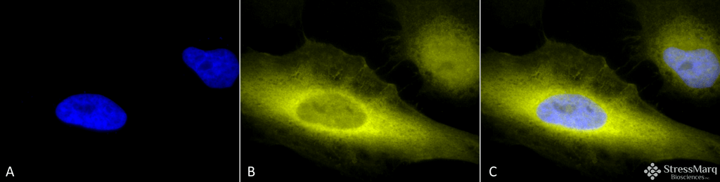
Immunocytochemistry/Immunofluorescence analysis using Rabbit Anti-Ubiquitin Polyclonal Antibody (SPC-119). Tissue: Heat Shocked Cervical cancer cell line (HeLa). Species: Human. Fixation: 2% Formaldehyde for 20 min at RT. Primary Antibody: Rabbit Anti-Ubiquitin Polyclonal Antibody (SPC-119) at 1:100 for 12 hours at 4°C. Secondary Antibody: R-PE Goat Anti-Rabbit (yellow) at 1:200 for 2 hours at RT. Counterstain: DAPI (blue) nuclear stain at 1:40000 for 2 hours at RT. Localization: Cytoplasm. Magnification: 100x. (A) DAPI (blue) nuclear stain. (B) Anti-Ubiquitin Antibody. (C) Composite. Heat Shocked at 42°C for 1h.
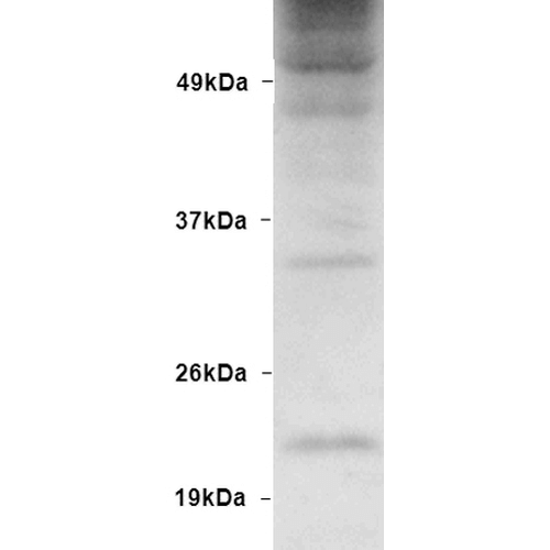
Western blot analysis of Human Embryonic kidney epithelial cell line (HEK293) lysate showing detection of Ubiquitin protein using Rabbit Anti-Ubiquitin Polyclonal Antibody (SPC-119). Primary Antibody: Rabbit Anti-Ubiquitin Polyclonal Antibody (SPC-119) at 1:1000.
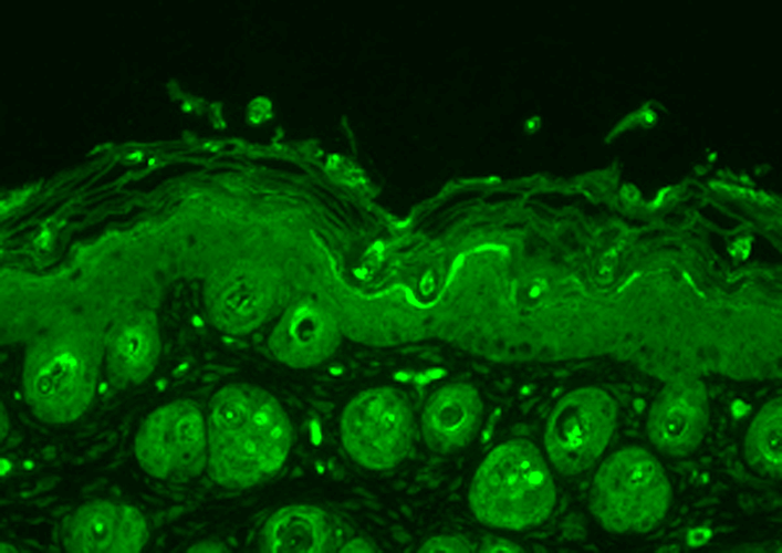
Immunohistochemistry analysis using Rabbit Anti-Ubiquitin Polyclonal Antibody (SPC-119). Tissue: backskin. Species: Mouse. Fixation: Bouin’s Fixative Solution. Primary Antibody: Rabbit Anti-Ubiquitin Polyclonal Antibody (SPC-119) at 1:100 for 1 hour at RT. Secondary Antibody: FITC Goat Anti-Rabbit (green) at 1:50 for 1 hour at RT. Localization: Cytoplasm.
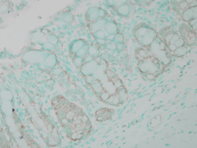
Immunohistochemistry analysis using Rabbit Anti-Ubiquitin Polyclonal Antibody (SPC-119). Tissue: colon carcinoma. Species: Human. Fixation: Formalin. Primary Antibody: Rabbit Anti-Ubiquitin Polyclonal Antibody (SPC-119) at 1:100000 for 12 hours at 4°C. Secondary Antibody: Biotin Goat Anti-Rabbit at 1:2000 for 1 hour at RT. Counterstain: Methyl Green at 200uL for 2 min at RT. This image was produced using an amplifying IHC wash buffer. The antibody has therefore been diluted more than is recommended for other applications..
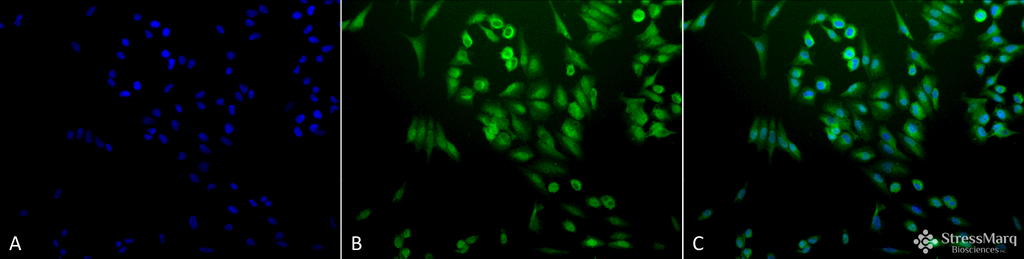
Immunocytochemistry/Immunofluorescence analysis using Rabbit Anti-Ubiquitin Polyclonal Antibody (SPC-119). Tissue: Cervical cancer cell line (HeLa). Species: Human. Fixation: 2% Formaldehyde for 20 min at RT. Primary Antibody: Rabbit Anti-Ubiquitin Polyclonal Antibody (SPC-119) at 1:200 for 12 hours at 4°C. Secondary Antibody: FITC Goat Anti-Rabbit (green) at 1:200 for 2 hours at RT. Counterstain: DAPI (blue) nuclear stain at 1:40000 for 2 hours at RT. Localization: Cytoplasm. Magnification: 20x. (A) DAPI (blue) nuclear stain. (B) Anti-Ubiquitin Antibody. (C) Composite.
 Powered by Bioz
StressMarq Biosciences :
Based on validation through cited publications.