| Product Name | Glutathione S-Transferase Activity Kit (Discontinued) |
| Description |
Quantitative fluorometric measurement of the activity of GST |
| Platform | Microplate |
| Sample Types | Cell lysates, EDTA Plasma, Heparin Plasma, Serum, Urine |
| Detection Method | Fluorometric Assay |
| Assay Type | Direct Enzyme Activity Assay |
| Utility | Fluorometric assay used to quantitatively measure the activity of glutathione S-transferase in samples. |
| Precision | Intra Assay Precision:Four serum samples were diluted 1:2 with Assay Buffer and run in replicates of 16 in an assay. The mean and precision of the calculated GST activities were: Sample 1- 315.9 mU/mL, 4.6% CV Sample 2- 221.2 mU/mL, 5.6% CV Sample 3- 88.2 mU/mL, 4.2% CV Sample 4- 22.7 mU/mL, 6.6% CV Inter Assay Precision: Four serum samples were diluted 1:2 with Assay Buffer and run in duplicates in twenty assays over multiple days by four operators. The mean and precision of the calculated GST activities were: Sample 1- 291.7 mU/mL, 12.6% CV Sample 2- 218.5 mU/mL, 11.0% CV Sample 3- 89.6 mU/mL, 10.4% CV Sample 4- 23.0 mU/mL, 15.9% CV |
| Incubation Time | 30 minutes |
| Number of Samples | 40 samples in duplicate |
| Other Resources | Kit Booklet , MSDS |
| Field of Use | Not for use in humans. Not for use in diagnostics or therapeutics. For in vitro research use only. |
Properties
| Storage Temperature | 4ºC | |||||||||||||||||||||
| Shipping Temperature | Blue Ice | |||||||||||||||||||||
| Product Type | Activity Kits | |||||||||||||||||||||
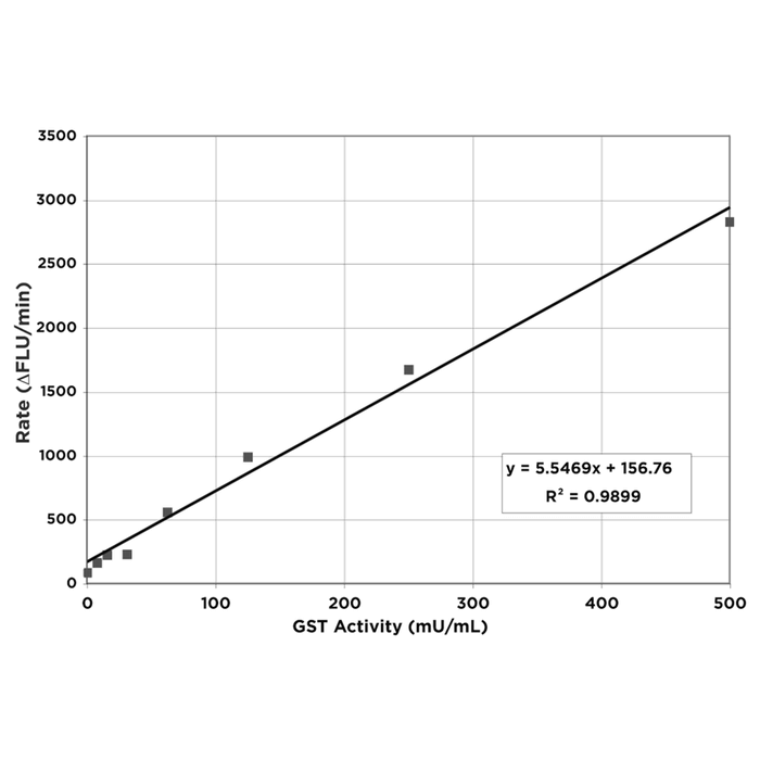
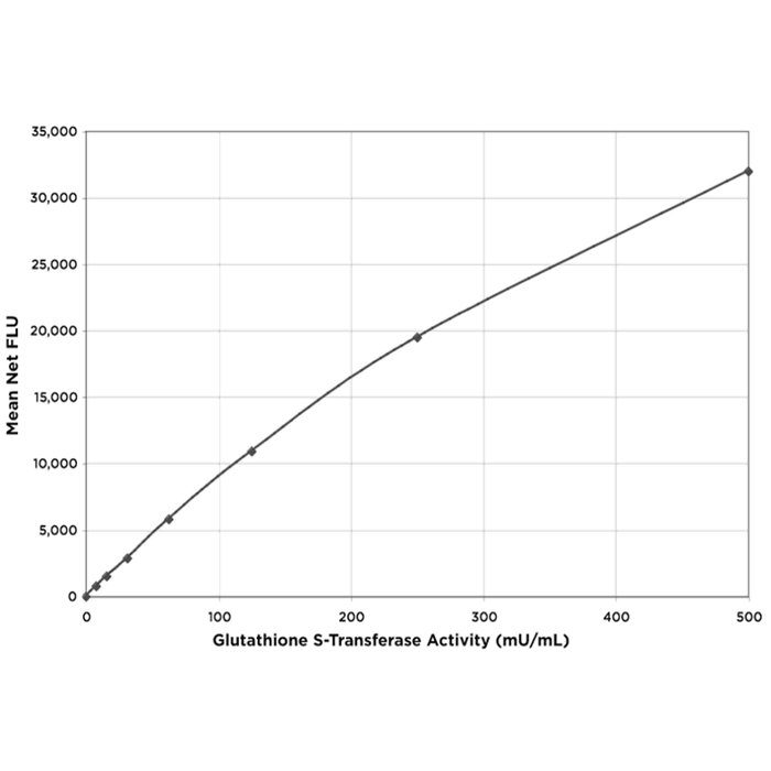
| Assay Overview | The Glutathione S-Transferase Activity kit is designed to quantitatively measure the activity of GST present in a variety of samples. A GST standard is provided to generate a standard curve for the assay and all samples should be read off the standard curve. The kit utilizes a non-fluorescent molecule that is a substrate for the GST enzyme which covalently attaches to glutathione (GSH) to yield a highly fluorescent product. Mixing the sample or standard with the supplied Detection Reagent and GSH and incubating at room temperature for 30 minutes yields a fluorescent product which is read at 460 nm in a fluorescent plate reader with excitation at 390 nm. The activity of the GST in the sample is calculated, after making a suitable correction for any dilution of the sample, using software available with most fluorescence plate readers. We have provided a 96 well plate for measurement but this assay is adaptable for higher density plate formats. The end user should ensure that their HTS black plate is suitable for use with these reagents prior to running samples. | |||||||||||||||||||||
| Kit Overview |
|
|||||||||||||||||||||
| Cite This Product | Glutathione S-Transferase Activity Kit (StressMarq Biosciences Inc., Victoria BC CANADA, Catalog # SKT-203) |
Biological Description
| Alternative Names | γ-L-Glutamyl-L-cysteinylglycine Activity Kit (2S)-2-Amino-5-[[(2R)-1-(carboxymethylamino)-1-oxo- 3-sulfanylpropan-2-yl]amino]-5-oxopentanoic acid Activity Kit |
| Research Areas | Cancer, Oxidative Stress |
| Scientific Background |
The Glutathione S-Transferase (GST) family of isozymes function to detoxify and neutralize a wide variety of electrophilic molecules by mediating their conjugation with reduced glutathione (1). Human GSTs are encoded by 5 gene families, expressing in almost all tissues as four cytosolic and one microsomal forms. Dividing the family by isoelectric points, the basic alpha (pI 8–11), the neutral mu (pI 5–7) and acidic pi (pH<5) classes are populated by additional subclasses, each isozyme displaying differential specificity for given electrophilic molecules (2). Given its pivotal role in ameliorating oxidative stress/damage, GST activity has been repeatedly investigated as a biomarker for arthritis, asthma, COPD, and multiple forms of cancer, as well as an environmental marker (3-7). Examination of GST isoforms and activity in human cancers, tumors and tumor cell lines has revealed the predominance of the acidic pi class. Furthermore, this activity is thought to substantially contribute to the innate or acquired resistance of specific neoplasms to anticancer therapy (8,9). |
| References |
1. Habig, W, et al. J. Biol.Chem. 1974 249(22):7130-7139. 2. Cook, JA, et al. Cancer Res. 1991 51:1606-1612. 3. Dalle-Donne, I, et al. 2006 52(4):601-623. 4. Surapneni, KM & VSC Gopan. Ind.J.Clin.Biochem. 2008 23(1):41.44. 5. Mohan, SK & O Venkataramana. Ind.J.Med. Sci. 2007 61:9-14. 6. Ferrandina, G., et al. Ann. Oncol. 1997 8:343-350. 7. Otitoju, O & Onwarah, INE. African J. Biotech. 2007 6(12)1455-1459. 8. Shea, TC, et al. Cancer Res. 1988 48:527-533. 9. Shea, TC, et al. Cancer Res. 1990 50:6848-6853. |

Reviews
There are no reviews yet.