Discovery through Partnership | Excellence through Quality


Properties
| Storage Buffer | PBS pH7.4, 50% glycerol, 0.09% sodium azide *Storage buffer may change when conjugated |
| Storage Temperature | -20ºC, Conjugated antibodies should be stored according to the product label |
| Shipping Temperature | Blue Ice or 4ºC |
| Purification | Protein G Purified |
| Clonality | Monoclonal |
| Clone Number | Map.ERP57 |
| Isotype | IgG1 |
| Specificity | Detects ~57kDa |
| Cite This Product | StressMarq Biosciences Cat# SMC-168, RRID: AB_2163133 |
| Certificate of Analysis | 0.5 µg/ml of SMC-168 was sufficient for detection of ERp57 in 10 µg of heat shock Heal Lysate by colorimetric immunoblot analysis using Goat anti-mouse IgG:HRP as the secondary antibody. |
Biological Description
| Alternative Names | ERp60 Antibody, ERp61 Antibody, Grp57 Antibody, Grp58 Antibody, P58 Antibody, PDIA3 Antibody, PI PLC Antibody, 58 kDa glucose regulated protein antibody, 58 kDa glucose-regulated protein antibody, 58 kDa microsomal protein antibody, Disulfide isomerase ER 60 antibody, Disulfide isomerase ER-60 antibody, Endoplasmic reticulum resident protein 57 antibody, Endoplasmic reticulum resident protein 60 antibody, ER p57 antibody, ER protein 57 antibody, ER protein 60 antibody, ERp 57 antibody, ERp57 antibody, Glucose Regulated Protein 58 Kd antibody, GRP 57 antibody, GRP 58 antibody, GRP57 antibody, HsT17083 antibody, p58 antibody, PDIA 3 antibody, PDIA3 antibody, PDIA3_HUMAN antibody, Phospholipase C alpha antibody, PI PLC antibody, Protein disulfide isomerase A3 antibody, Protein disulfide isomerase family A member 3 antibody, Protein disulfide-isomerase A3 antibody |
| Research Areas | Cell Signaling, ER Proteins, Protein Trafficking |
| Cellular Localization | Endoplasmic Reticulum, Endoplasmic reticulum lumen, Melanosome |
| Accession Number | NP_005304.3 |
| Gene ID | 2923 |
| Swiss Prot | P30101 |
| Scientific Background | ERp57, also known as Glucose Regulated Protein 58 (Grp58), Hormone-Induced Protein-70 (HIP-70) and microsomal Carnitine Palmitoyltransferase, is a member of the protein disulfide isomerase family, containing two canonical CXHC tetrapeptide active site motifs (1-5). It has quite a few diverse roles. It functions as an accessory oxidoreductase involved in disulfide bond formation. In the ER, ERp57 interacts with membrane bound calnexin and soluble calreticulin (lectin chaperones) via their praline rich P-domain arms. Lectin chaperones bind nascent non-native glycoproteins, and position ERp57 to act upon the immature or misfolded glycoproteins that possess mono-glycosylated side chains. ERp57 deletion impairs posttranslational phases of influenza hema-glutinin folding, and causes accelerated release of MHC-I molecules, resulting in the coupling of sub-optimal peptides and reduced expression and stability on the cell surface (6). ERp57 also contains two thioredoxin active-site sequences, CGHC and an estrogen-binding domain. ERp57 is induced by both estrogen and leuteinizing-hormone-releasing hormone in the hippocampus (7). |
| References |
1. Herbert D.N. and Molinari M. (2007) Physiol Rev. 87: 1377-1408. 2. Williams D.B. (2005) J Cell Sci. 119: 615-623 3. Maattanen P., et al. (2006) Biochem Cell Biol. 84: 881-889. 4. Oliver J.D., et al. (1999) Mol Bio Cell. 10: 2573-2582. 5. Oliver J.D., et al. (1997) Science 275: 86-88. 6. Solda T., et al. (2006) J Biol Chem 281: 6219-6226. 7. Kimura T., et al. (2005) Biochem Biophys Research Communications. 331 (1): 224-230. 8. Chen, G., et al. (2002) Clin Cancer Res 8(7): 2298-2305. 9. Tan, P., et al. F. (2002) J Immunol 168(4): 1950-1960. |
Product Images
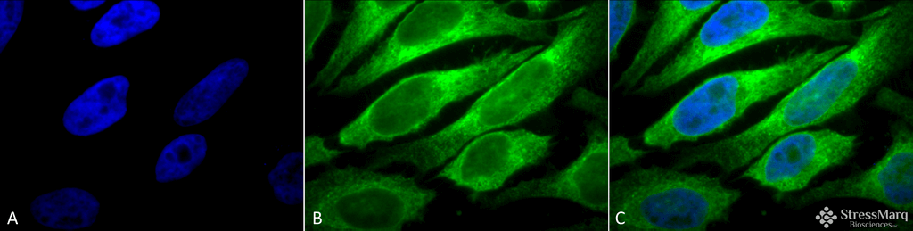
Immunocytochemistry/Immunofluorescence analysis using Mouse Anti-Erp57 (Grp58) Monoclonal Antibody, Clone Map.ERP57 (SMC-168). Tissue: Heat Shocked cervical cancer cells (HeLa). Species: Human. Fixation: 2% Formaldehyde for 20 min at RT. Primary Antibody: Mouse Anti-Erp57 (Grp58) Monoclonal Antibody (SMC-168) at 1:100 for 12 hours at 4°C. Secondary Antibody: FITC Goat Anti-Mouse (green) at 1:200 for 2 hours at RT. Counterstain: DAPI (blue) nuclear stain at 1:40000 for 2 hours at RT. Localization: Endoplasmic reticulum lumen. Melanosome. Magnification: 100x. (A) DAPI (blue) nuclear stain. (B) Anti-Erp57 (Grp58) Antibody. (C) Composite. Heat Shocked at 42°C for 1h.
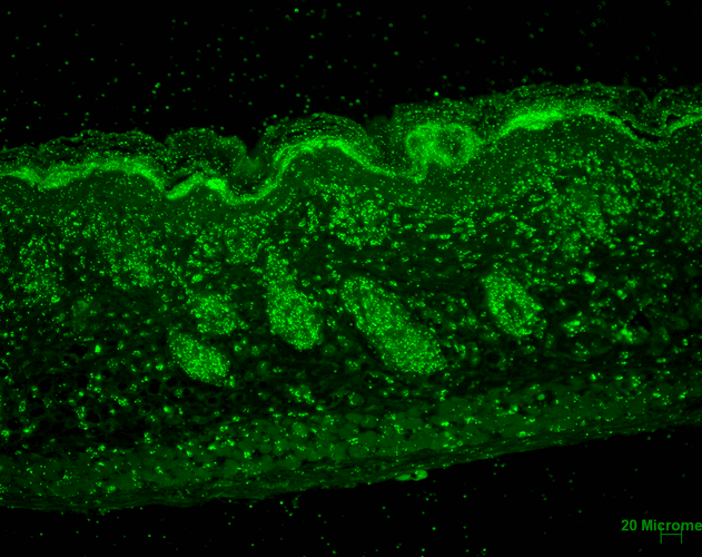
Immunohistochemistry analysis using Mouse Anti-Erp57 Monoclonal Antibody, Clone Map.ERP57 (SMC-168). Tissue: backskin. Species: Mouse. Fixation: Bouin’s Fixative and paraffin-embedded. Primary Antibody: Mouse Anti-Erp57 Monoclonal Antibody (SMC-168) at 1:100 for 1 hour at RT. Secondary Antibody: FITC Goat Anti-Mouse (green) at 1:50 for 1 hour at RT. Localization: Epidermis and Hair Follicles.
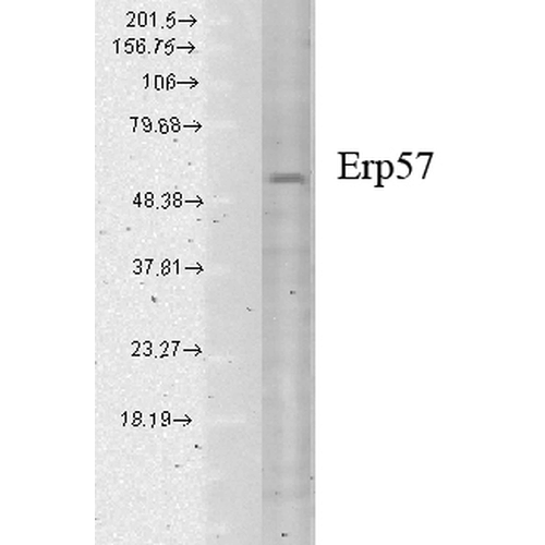
Western Blot analysis of Human cell lysates showing detection of Erp57 protein using Mouse Anti-Erp57 Monoclonal Antibody, Clone Map.ERP57 (SMC-168). Load: 15 µg. Block: 1.5% BSA for 30 minutes at RT. Primary Antibody: Mouse Anti-Erp57 Monoclonal Antibody (SMC-168) at 1:1000 for 2 hours at RT. Secondary Antibody: Sheep Anti-Mouse IgG: HRP for 1 hour at RT.
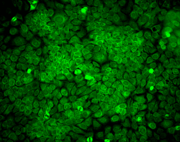
Immunocytochemistry/Immunofluorescence analysis using Mouse Anti-Erp57 Monoclonal Antibody, Clone Map.ERP57 (SMC-168). Tissue: HaCaT cells. Species: Human. Fixation: Cold 100% methanol for 10 minutes at -20°C. Primary Antibody: Mouse Anti-Erp57 Monoclonal Antibody (SMC-168) at 1:100 for 1 hour at RT. Secondary Antibody: FITC Goat Anti-Mouse (green) at 1:50 for 1 hour at RT. Localization: Cytoplasmic and perinuclear staining.
 Powered by Bioz
StressMarq Biosciences :
Based on validation through cited publications.