Wanida Vongpraseuth | April 24th, 2026 | Product Citation Reviews
The microtubule-associated protein tau, encoded by the MAPT gene, is a key regulator of neuronal structure and axonal transport in the brain. Alternative splicing of MAPT gives rise to multiple tau isoforms that differ in the number of microtubule-binding repeats and N-terminal inserts, contributing to functional diversity across neuronal contexts. Under pathological conditions, tau undergoes aberrant post-translational modifications (PTMs), including hyperphosphorylation and proteolytic cleavage, which promote conformational instability, misfolding, and subsequent aggregation.
In disease states, misfolded tau assembles into paired helical filaments (PHFs) that accumulate as insoluble neurofibrillary tangles (NFTs), a defining neuropathological feature of tauopathies such as Alzheimer’s disease (AD) and Chronic Traumatic Encephalopathy (CTE). Although intracellular tau aggregation has been extensively characterized, accumulating evidence highlights a significant role for extracellular tau species in propelling disease progression. Tau has now been shown to be capable of intercellular prion-like propagation, whereby extracellular tau seeds template misfolding and aggregation in recipient neurons. Moreover, soluble extracellular tau fragments have been implicated in direct synaptic dysfunction, where they impair synaptic plasticity – the activity-dependent strengthening and remodeling of synaptic connections that underlies learning and memory.
Targeting extracellular tau
Efforts to develop tau-targeting therapeutics have culminated in the development of next-generation antibodies designed to neutralize extracellular tau species. However, clinical trials of antibodies directed against the N-terminal region have thus far shown limited efficacy, prompting a strategic shift toward alternative epitopes. In particular, increasing attention has focused on the microtubule-binding region (MTBR) and the adjacent pseudorepeat R’ (MTBR/R’), which are highly enriched in aggregation-prone tau fragments.
Notably, levels of tau species containing the MTBR/R’ region are elevated in Alzheimer’s disease (AD) brains and are strongly associated with tau’s intrinsic propensity to aggregate. In an international collaboration involving Trinity College Dublin, Zhengzhou University, Talisman Therapeutics, and the Ann Romney Center for Neurologic Diseases, researchers investigated this region as a candidate synaptotoxic domain. The study examined its effects on long-term potentiation (LTP), a well-established electrophysiological measure of synaptic plasticity that reflects the strength of synaptic transmission between neurons. In parallel, the investigators evaluated two novel antibodies targeting the MTBR/R’ region to determine their ability to mitigate tau-induced synaptic dysfunction.
Synaptotoxic tau fragments
Ondrejcak et al. investigated the contribution of extracellular tau species to synaptic dysfunction by analyzing secretomes, the full complement of proteins released by cells, from induced pluripotent stem cell-derived neurons (iNs). These neurons were generated from individuals with trisomy 21 (Ts21), the genetic basis of Down syndrome, a condition strongly associated with early-onset Alzheimer’s disease.
To assess functional effects in vivo, these neuronal secretomes were introduced into the brains of anesthetized rats. Subsequent electrophysiological recordings in the hippocampus revealed that Ts21-derived secretomes exhibited impaired LTP, a well-established correlate of synaptic plasticity and memory formation. Further proteomic analysis using mass spectrometry revealed that these secretomes were further enriched in tau fragments containing the microtubule-binding region and adjacent pseudorepeat R’ (MTBR/R’), implicating this domain in synaptotoxic activity.
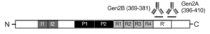
Figure 1. Tau subunit structure with antibody binding sites of Gen2A and Gen2B shown. From left to right: N-terminus, N-terminal Projection Domain, N1 and N2 inserts (I1/I2), Proline-Rich Region (P1/P2), Microtubule-Binding Domain MTBR (R1-R4), Pseudorepeat (R’), C-terminus. Figure taken from Ondrejcak et al., 2025 used under license CC BY 4.0).
Critically, immunodepletion experiments demonstrated that removal of MTBR/R’-containing tau species abolished the LTP deficit and restored normal synaptic function. This was achieved using antibodies directed against the MTBR/R’ region, including a commercially available Tau5 antibody (which recognizes the P2 domain) and two newly generated monoclonal antibodies, Gen2A and Gen2B. Together, these antibodies collectively span tau residues 218 to 410. In contrast, Tau46, an antibody targeting the distal C-terminal region of tau, failed to rescue synaptic impairment, underscoring the specificity of the MTBR/R’ domain in mediating toxicity.
The dGAE region
To further define the tau species responsible for synaptotoxicity, the investigators fractionated Ts21 neuronal secretomes using size-exclusion chromatography (SEC), separating the material into more than 20 discrete fractions according to molecular size. Functional screening of these fractions showed that fractions 15 and 16 contained the highest abundance of MTBR-containing tau species and, importantly, produced robust inhibition of LTP when injected into the hippocampus of anesthetized rats.
This synaptic impairment was effectively prevented by co-administration of the Gen2A antibody, indicating that the toxic activity of these fractions was specifically mediated by MTBR/R’-containing tau species. To determine whether this effect could be recapitulated using defined recombinant tau fragments, authors utilized StressMarq’s Tau dGAE (297-391) Monomers (catalog# SPR-444), which encompass the core MTBR/R’ region. Notably, even in the absence of pre-aggregation, this monomeric fragment reproduced the LTP deficits at physiologically relevant concentrations.
Rescuing synaptic plasticity
Researchers next examined whether MTBR/R’-containing tau species identified in their experimental system are also present in human AD brain tissue. Aqueous extracts prepared from two post-mortem AD patient brains (AD1 and AD6) were analyzed using MTBR/R’-specific antibodies. Both Gen2A and Gen2B robustly detected tau fragments containing the MTBR/R’ region, confirming the presence of these species in human disease-derived material.
To assess their functional relevance, the authors injected the AD brain extracts into the hippocampus of anesthetized rats and recorded synaptic activity. These extracts reliably disrupted long-term potentiation, indicating impaired synaptic plasticity. Importantly, co-injection with either Gen2A or Gen2B preserved normal LTP, whereas control IgG had no protective effect. These findings support the conclusion that MTBR/R’-containing tau fragments present in AD brain extracts are directly responsible for the observed synaptic deficits and can be selectively neutralized by targeted antibodies.
A particularly compelling series of experiments examined whether antibody treatment could reverse established synaptic dysfunction. Following injection of AD1 brain extract, human tau species remained detectable in the rat hippocampus for weeks, and LTP remained significantly suppressed. Remarkably, a single administration of anti-tau antibodies, including Tau5, Gen2A, or Gen2B, rapidly restored robust LTP within minutes of treatment. In contrast, control IgG had no effect on synaptic function.
Summary
This study provides compelling evidence that extracellular tau fragments containing the MTBR/R′ domain are key mediators of synaptic toxicity in AD. The experimental data demonstrate that tau species spanning approximately amino acids 218-410 are both necessary and sufficient to impair long-term potentiation in vivo across multiple patient-derived and recombinant preparations. Functionally, these fragments disrupt synaptic plasticity, thereby compromising processes essential for learning and memory.
Importantly, the study further demonstrates that MTBR/R′-directed antibodies can rapidly and effectively neutralize these toxic tau species. In addition to preventing synaptic impairment, antibody treatment was able to reverse established LTP deficits even after pathology had been introduced, indicating a capacity to restore synaptic function rather than merely prevent damage.
Together, these findings support a model in which MTBR/R′-containing extracellular tau fragments act as proximal drivers of synaptic dysfunction in Alzheimer’s disease, while also highlighting their potential as therapeutically tractable targets for both disease modification and functional recovery.
Related StressMarq products
StressMarq manufactures a wide range of neurodegenerative proteins in different conformations, providing researchers with high-quality tools designed to support experimental reproducibility and biological relevance in models of protein misfolding, aggregation, and prion-like propagation. Visit our website for more information, including the latest scientific publications using our specialized tau, amyloid beta and alpha synuclein proteins for neurodegenerative disease research. Of particular relevance are the Tau dGAE (297-391) AD-mimic Pre-formed Fibrils (catalog# SPR-502), which replicate the characteristic filament fold observed in Alzheimer’s disease-derived tau aggregates. This model system provides a structurally relevant tool for studying aggregation and seeding mechanisms associated with MTBR/R′-containing tau species.
References
- The role of tau in neuronal function and neurodegeneration. Aranda-Abreu, G. E. et al. Neurol Int. 2025.
- Synaptotoxic effects of extracellular tau are mediated by its microtubule-binding region. Ondrejcak, T. et al. Acta Neuropathol. 2025.

Leave a Reply