Properties
| Storage Buffer | PBS pH7.4, 50% glycerol, 0.09% sodium azide *Storage buffer may change when conjugated |
| Storage Temperature | -20ºC, Conjugated antibodies should be stored according to the product label |
| Shipping Temperature | Blue Ice or 4ºC |
| Purification | Protein G Purified |
| Clonality | Monoclonal |
| Clone Number | N327A/38 (Formerly sold as S327A-38) |
| Isotype | IgG2b |
| Specificity | Detects ~170kDa. Does not react with NR2B. |
| Cite This Product | StressMarq Biosciences Cat# SMC-434, RRID: AB_2701559 |
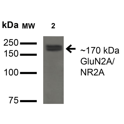
| Certificate of Analysis | 1 µg/ml of SMC-434 was sufficient for detection of GluN2A/NR2A in 20 µg of COS cells transiently transfected with GFP-tagged NR2A lysate by colorimetric immunoblot analysis using Goat anti-mouse IgG:HRP as the secondary antibody. |
Biological Description
| Alternative Names | NMDA 2A Antibody, NMDAR2A Antibody, NMDAR 2A Antibody, NMDA Receptor 2A Antibody, Glutamate Receptor Antibody, GRIN2A Antibody, Glutamate [NMDA] Receptor subunit epsilon-1 Antibody, Glutamate receptor ionotropic N methyl D aspartate 2A Antibody, HNR2A Antibody, N methyl D aspartate receptor channel Antibody, subunit epsilon 1 Antibody, N Methyl D Aspartate Receptor Subtype 2A Antibody, N methyl D aspartate receptor subunit 2A Antibody, NMDA receptor subtype 2A Antibody, NMDA Receptor Type 2A Antibody, OTTHUMP00000160135 Antibody, OTTHUMP00000174531 Antibody |
| Research Areas | Cell Signaling, Glutamate Receptors, Neuroscience, Neurotransmitter Receptors, NMDA Receptors |
| Cellular Localization | Cell Junction, Cell membrane |
| Accession Number | NP_036705.3 |
| Gene ID | 24409 |
| Swiss Prot | Q00959 |
| Scientific Background | N-methyl-D-aspartate (NMDA) receptors are a class of ionotropic glutamate-gated ion channels. These receptors have been shown to be involved in long-term potentiation, an activity-dependent increase in the efficiency of synaptic transmission thought to underlie certain kinds of memory and learning. NMDA receptor channels are heteromers composed of the key receptor subunit NMDAR1 (GRIN1) and 1 or more of the 4 NMDAR2 subunits: NMDAR2A (GRIN2A), NMDAR2B (GRIN2B), NMDAR2C (GRIN2C) and NMDAR2D (GRIN2D). |
| References | 1. Teng H.J., et al. (2010) PLoS ONE. 5: e13342. |
Product Images

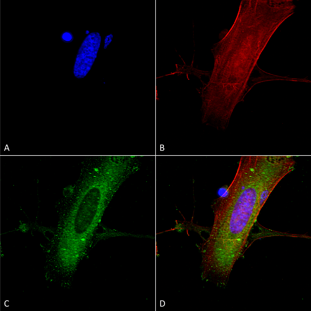
Immunocytochemistry/Immunofluorescence analysis using Mouse Anti-GluN2A/NR2A Monoclonal Antibody, Clone N327A/38 (SMC-434). Tissue: Neuroblastoma cells (SH-SY5Y). Species: Human. Fixation: 4% PFA for 15 min. Primary Antibody: Mouse Anti-GluN2A/NR2A Monoclonal Antibody (SMC-434) at 1:100 for overnight at 4°C with slow rocking. Secondary Antibody: AlexaFluor 488 at 1:1000 for 1 hour at RT. Counterstain: Phalloidin-iFluor 647 (red) F-Actin stain; Hoechst (blue) nuclear stain at 1:800, 1.6mM for 20 min at RT. (A) Hoechst (blue) nuclear stain. (B) Phalloidin-iFluor 647 (red) F-Actin stain. (C) GluN2A/NR2A Antibody (D) Composite.

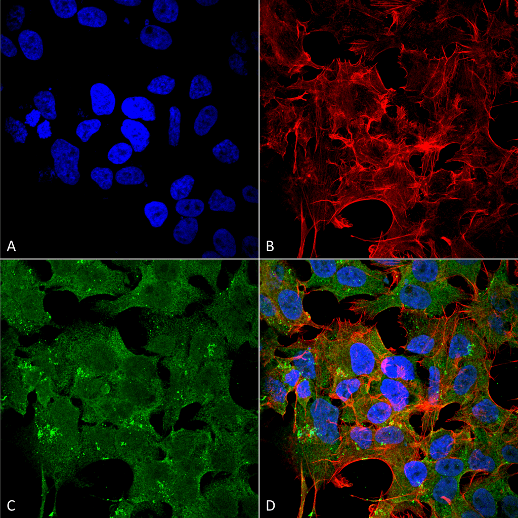
Immunocytochemistry/Immunofluorescence analysis using Mouse Anti-GluN2A/NR2A Monoclonal Antibody, Clone N327A/38 (SMC-434). Tissue: Neuroblastoma cell line (SK-N-BE). Species: Human. Fixation: 4% Formaldehyde for 15 min at RT. Primary Antibody: Mouse Anti-GluN2A/NR2A Monoclonal Antibody (SMC-434) at 1:100 for 60 min at RT. Secondary Antibody: Goat Anti-Mouse ATTO 488 at 1:200 for 60 min at RT. Counterstain: Phalloidin Texas Red F-Actin stain; DAPI (blue) nuclear stain at 1:1000, 1:5000 for 60 min at RT, 5 min at RT. Localization: Cell Membrane, Cytoplasm. Magnification: 60X. (A) Phalloidin Texas Red F-Actin stain; DAPI (blue) nuclear stain. (B) Anti-GluN2A/NR2A Antibody. (C) Composite. (A) DAPI (blue) nuclear stain. (B) Phalloidin Texas Red F-Actin stain. (C) GluN2A/NR2A Antibody. (D) Composite.

Western Blot analysis of Monkey COS cells transfected with GFP-tagged NR2A showing detection of ~170 kDa GluN2A/NR2A protein using Mouse Anti-GluN2A/NR2A Monoclonal Antibody, Clone N327A/38 (SMC-434). Lane 1: Molecular Weight Ladder. Lane 2: Monkey COS cells transfected with GFP-tagged NR2A. Load: 15 µg. Block: 2% BSA and 2% Skim Milk in 1X TBST. Primary Antibody: Mouse Anti-GluN2A/NR2A Monoclonal Antibody (SMC-434) at 1:200 for 16 hours at 4°C. Secondary Antibody: Goat Anti-Mouse IgG: HRP at 1:1000 for 1 hour RT. Color Development: ECL solution for 6 min in RT. Predicted/Observed Size: ~170 kDa.


Reviews
There are no reviews yet.