Discovery through Partnership | Excellence through Quality


Properties
| Storage Buffer | PBS pH7.2, 50% glycerol, 0.09% sodium azide *Storage buffer may change when conjugated |
| Storage Temperature | -20ºC, Conjugated antibodies should be stored according to the product label |
| Shipping Temperature | Blue Ice or 4ºC |
| Purification | Protein G Purified |
| Clonality | Monoclonal |
| Clone Number | PIN.1 |
| Isotype | IgG1 |
| Specificity | Detects ~33-35kDa protein doublet corresponding to the molecular mass of the p33 and p35 forms of human CD74. |
| Cite This Product | StressMarq Biosciences Cat# SMC-116, RRID: AB_2260133 |
| Certificate of Analysis | 1 µg/ml of SMC-116 was sufficient for detection of CD74 in 20 µg of PALA cell lysates by colorimetric immunolot analysis using goat anti-mouse IgG: AP as the secondary antibody. |
Biological Description
| Alternative Names | DHLAG Antibody, HLA DR gamma Antibody, HLADG Antibody, p33 Antibody, p35 Antibody, Protein 41 Antibody, CD 74 antibody, CD74 antibody, CD74 antigen (invariant polypeptide of major histocompatibility complex, class II antigen-associated) antibody, CD74 antigen antibody, CD74 molecule antibody, CD74 molecule, major histocompatibility complex, class II invariant chain antibody, CLIP antibody, DHLAG antibody, Gamma chain of class II antigens antibody, HG2A_HUMAN antibody, HLA class II histocompatibility antigen gamma chain antibody, HLA DR antigens associated invariant chain antibody, HLA DR gamma antibody, HLA-DR antigens-associated invariant chain antibody, HLA-DR-gamma antibody, HLADG antibody, HLADR antigens associated invariant chain antibody Ia antigen associated invariant chain antibody, Ia antigen-associated invariant chain antibody, Ia associated invariant chain antibody, Ia gamma antibody, Ii antibody, Invariant polypeptide of major histocompatibility complex class II antigen associated antibody, la-gamma antibody, Major histocompatibility complex class II invariant chain antibody, MHC HLA DR gamma chain antibody, MHC HLA-DR gamma chain antibody, p33 antibody, p35 antibody, Protein 41 antibody |
| Research Areas | Adaptive Immunity, B Cell CD Markers, B Cells, Immunology |
| Cellular Localization | Cell membrane, Endoplasmic Reticulum, Endoplasmic reticulum membrane, Endosome, Golgi apparatus, Lysosome |
| Accession Number | NP_001020329.1 |
| Gene ID | 972 |
| Swiss Prot | P04233 |
| Scientific Background | CD74 is a non-polymorphic type II integral membrane protein. It has a short N-terminal cytoplasmic tail of 28 amino acids, followed by a single 24-aa transmembrane region and an approximately 150-aa lumenal domain (1). The CD74 chain is thought to function mainly as an MHC class II chaperone, which promotes ER exit of MHC class II molecules, directs them to endocytic compartments, prevents peptide binding in the ER, and contributes to peptide editing in the MHC class II compartment. Class II MHC and Ii expression was believed to be restricted to classical antigen-presenting cells (APC); however, during inflammation, other cell types, including mucosal epithelial cells, have also been reported to express class II MHC molecules (2). Experiments that investigate cell-surface CD74 are complicated by the fact that CD74 remains on the cell surface for a very short time. The surface half-life of CD74 was calculated to be fewer than 10 minutes (3). CD74 however has also recently been shown to have a role as an accessory-signaling molecule because of its high-affinity binding to the pro-inflammatory cytokine, macrophage migration-inhibitory factor (MIF) (3). The restricted expression of CD74 by normal tissues and its very rapid internalization make CD74 an attractive therapeutic target for both cancer and immunologic diseases (4). |
| References |
1. Becker-Hermann, S., Arie, G., Medvedovsky H, Kerem A, and Shachar I. (2005) Mol Bio Cell. 16(11):5061-9. 2. Barrera CA., et al (2005) J Histochem Cytochem 53 (12): 1481-9. 3. Starlets D., et al. (2006) Blood. 107 (12): 4807-4816. 4. Burton JD., et al. (2004). Clin Cancer Res. 10(19): 6606-11. 5. Denzin L.K., Hammond, C. and Cresswell, P. (1996) J. Exp. Med. 184: 2153-2165. 6. Denzin L.K., Robbins N.F., Carboy-Newcomb C. and Cresswell P. (1994) Immunity 1: 595-606. |
Product Images

Immunocytochemistry/Immunofluorescence analysis using Mouse Anti-CD74 Monoclonal Antibody, Clone PIN 1.1 (SMC-116). Tissue: Cervical cancer cell line (HeLa). Species: Human. Fixation: 2% Formaldehyde for 20 min at RT. Primary Antibody: Mouse Anti-CD74 Monoclonal Antibody (SMC-116) at 1:100 for 12 hours at 4°C. Secondary Antibody: FITC Goat Anti-Mouse (green) at 1:200 for 2 hours at RT. Counterstain: DAPI (blue) nuclear stain at 1:40000 for 2 hours at RT. Localization: Cell membrane. Endoplasmic reticulum membrane. Golgi apparatus. Endosome. Lysosome. Magnification: 100x. (A) DAPI (blue) nuclear stain. (B) Anti-CD74 Antibody. (C) Composite.
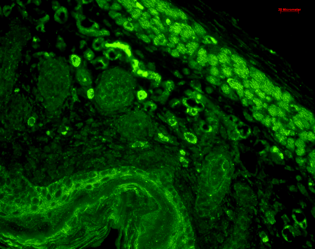
Immunohistochemistry analysis using Mouse Anti-CD74 Monoclonal Antibody, Clone PIN 1.1 (SMC-116). Tissue: backskin. Species: Mouse. Fixation: Bouin’s Fixative and paraffin-embedded. Primary Antibody: Mouse Anti-CD74 Monoclonal Antibody (SMC-116) at 1:100 for 1 hour at RT. Secondary Antibody: FITC Goat Anti-Mouse (green) at 1:50 for 1 hour at RT. Localization: Beautiful basal to suprabasal staining in epidermis, dermis, hair follicles and muscle.
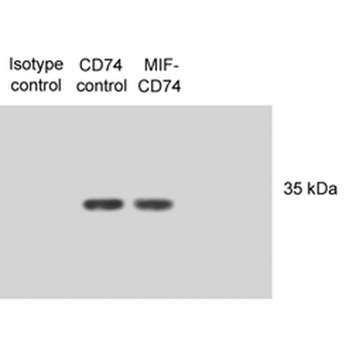
Western Blot analysis of Human N87 cell lysates showing detection of CD74 protein using Mouse Anti-CD74 Monoclonal Antibody, Clone PIN 1.1 (SMC-116). Primary Antibody: Mouse Anti-CD74 Monoclonal Antibody (SMC-116) at 1:1000. Lysates treated with macrophage inhibitory factor (MIF). Courtesy of: Victor E. Reyes, University of Texas Medical Branch, USA.
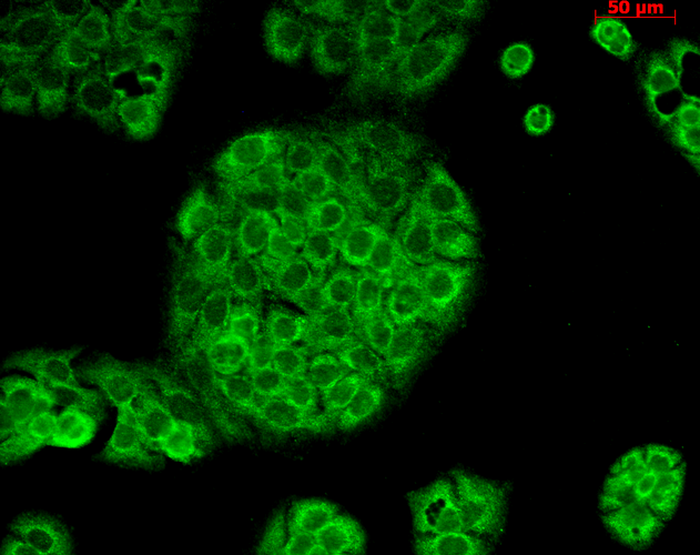
Immunocytochemistry/Immunofluorescence analysis using Mouse Anti-CD74 Monoclonal Antibody, Clone PIN 1.1 (SMC-116). Tissue: HaCaT cells. Species: Human. Fixation: Cold 100% methanol for 10 minutes at -20°C. Primary Antibody: Mouse Anti-CD74 Monoclonal Antibody (SMC-116) at 1:100 for 1 hour at RT. Secondary Antibody: FITC Goat Anti-Mouse (green) at 1:50 for 1 hour at RT. Localization: Cytoplasmic Staining.
 Powered by Bioz
StressMarq Biosciences :
Based on validation through cited publications.