

Antibody Genetics & Amyloid Beta Immunity in Alzheimer’s Disease
Alzheimer’s disease (AD), defined by the accumulation of amyloid beta plaques and tau pathology in the brain, is the most common cause of dementia worldwide. The immune system’s role in shaping disease progression has become an increasingly important area of investigation. Naturally produced auto-antibodies targeting amyloid beta have been shown to help clear amyloid beta plaques. Furthermore, therapeutic monoclonal antibodies that target amyloid beta have shown promise in the treatment of Alzheimer’s disease, however, effectiveness has varied, with potentially serious side effects observed in some cases.
Immunoglobulin GM (γ marker) allotypes are hereditary antigenic variants found in the constant region of IgG antibodies. GM allotypes have previously been identified as genetic risk factors for AD, yet why and how they influence risk has remained unclear. A study by Pandey et al. published in Human Immunology takes a significant step toward answering this question by examining whether GM allotypes affect naturally occurring antibody responses to amyloid beta 42, a central pathological peptide in AD. Amyloid beta is a 42-residue peptide which forms soluble oligomers, considered the most neurotoxic of the amyloid species. These oligomers can seed aggregation across brain regions and are known to disrupt neuronal function and signaling. This disruption leads to inflammation and neurodegeneration.
Study design: Genotyping and ELISA
Pandey and colleagues at the Medical University of South Carolina (MUSC) analyzed DNA and plasma samples from 100 clinically diagnosed AD patients and 100 cognitively normal controls. Clinical diagnoses were supported by cerebrospinal fluid (CSF) biomarkers that were available for most of the participants (amyloid beta 42, total tau, and phosphorylated tau). Two key components were measured during the study, GM and KM immunoglobulin allotypes (genetic variants of IgG and Igκ chains) and naturally occurring IgG antibodies against amyloid beta 42. These antibodies were quantified using an enzyme-linked immunosorbent assay (ELISA).
Genotyping of the samples included several GM allotypes (GM 3, GM 17, GM 23, GM 5, GM 2) and KM allotypes (KM 1 and KM 3). Plasma IgG antibodies against amyloid beta 42 were measured by ELISA on microplates coated with StressMarq’s Amyloid Beta 1-42 Oligomers (catalog #SPR-488). Diluted patient plasma was incubated on coated wells to permit specific antibody binding to the immobilized oligomers. Bound plasma IgG was detected with HRP-conjugated anti-human IgG secondary antibody, and the enzymatic signal was quantified colorimetrically.
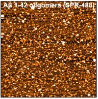
Figure 1: [Image from StressMarq Biosciences] Atomic force microscopy (AFM) of Amyloid Beta 1-42 Oligomers (catalog #SPR-488).
Experimental data supports clinical validity
The authors reported that several of the findings supported the classic Alzheimer’s disease signature, confirming the clinical validity of the cohort. APOE (apolipoprotein E) genotyping showed a higher frequency of the APOE ε4 allele in AD cases compared to controls. APOE ε4 is a genetic variant of APOE associated with a higher risk of developing AD. Additionally, phosphorylated tau and total tau levels in the CSF were elevated in AD patients versus controls. This is consistent with tau hyperphosphorylation and misfolding causing microtubule detachment and release of tau into the surrounding fluid. Meanwhile, significantly lower levels of amyloid beta 42 were observed in the CSF in AD patients compared to controls. This is a well-established biomarker of amyloid pathology where the peptide becomes insoluble and is deposited in brain plaques.
Specific allotypes affect amyloid beta immunity
Pandey et al. carried out further statistical analysis, combining their ELISA data on the levels of anti-amyloid beta 42 in a particular plasma sample and genotyping to reveal the specific allotypes of patients and controls. The analysis showed that GM 3/17 allotypes significantly influenced anti-amyloid beta 42 antibody levels in AD patients, but not in the cognitively normal control samples. Among the samples, GM 17/17 homozygotes had the lowest antibody levels, whereas GM 3/3 homozygotes had the highest.
Notably, increasing copies of the GM 17 allele correlated with reduced anti-amyloid beta 42 immunity (GM 3/3 > GM 3/17 > GM 17/17). A similar pattern for GM 23 allotype emerged, where significant effects were only seen in AD patients. GM 23 -/- individuals had the lowest antibody levels, whereas homozygous GM 23 +/+ had the highest (GM 23 −/− < GM 23 +/− < GM 23 +/+). There were no other significant associations for other GM or KM allotypes.
Mechanisms of GM allotype impact
The authors proposed several mechanisms as to why GM allotypes influence antibody responses to amyloid beta 42. Amino acid substitutions defining GM 3 and GM 23 may alter how membrane‑bound IgG on memory B cells recognizes and internalizes amyloid beta 42, enhancing antigen processing and presentation. Furthermore, GM allotypes reside in the constant region of IgG, but structural studies show that constant‑region changes can influence variable‑region conformation and antigen binding kinetics, thus improving antibody affinity via conformational effects.
Finally, GM 3 and GM 23 are in strong linkage disequilibrium, meaning these variants are often inherited together. It is therefore possible that their association with amyloid beta immunity reflects linkage disequilibrium with a currently unidentified immune-modulating gene. To explain why these GM-linked antibody responses were only seen in AD patients, the researchers suggested that in Alzheimer’s, amyloid beta 42 antibodies may recognize different epitopes to those in healthy individuals.
With therapeutic monoclonal amyloid beta antibodies only showing limited success, the development of amyloid beta vaccines remains an important but challenging area of research. Understanding the genetic and immunological factors that shape natural antibody responses to amyloid beta could help explain who benefits from immunotherapies and how to design safe, effective vaccines.
Summary
Despite its limitations regarding sample size and the single ethnicity of the subjects (Caucasian), this study provides the first evidence that suggests GM immunoglobulin allotypes (GM 3/17 and GM 23) may be associated with the level of naturally occurring anti‑amyloid beta 42 antibodies in patients with Alzheimer’s disease. If GM allotypes influence natural antibody responses, they may also influence vaccine responsiveness, therapeutic antibody efficacy, and disease progression rates. Incorporating GM allotype data into trial design could allow researchers to group participants more effectively and uncover genotype-linked differences in treatment response.
Related StressMarq products
StressMarq Biosciences offers highly validated research tools for neurodegenerative disease research. Our complete range of pathologically relevant amyloid beta proteins include monomers, oligomers and fibrils, such as Amyloid Beta Pyroglutamate 3-42 Pre-formed Fibrils (catalog #SPR-492). This truncated peptide replicates the b-sheet-rich structures found in amyloid plaques and is particularly neurotoxic due to its stability and ability to seed. For detection, amyloid beta antibodies (catalog #SPC-506, #SPC-507, #SPC-816, #SMC-618, #SMC-619, #SMC-620) are available with a selection of conjugates for ease of use across a range of assays.
References
- Immunoglobulin γ allotypes influence the level of auto-antibody responses to amyloid-β in patients with Alzheimer’s disease. Pandey, J.P. et al., Hum Immunol. 2025 Dec 9;87(1):111632.
Leave a Reply