
Cellular Mechanisms in Early Parkinson’s Disease
Parkinson’s disease (PD) is characterized by the accumulation of misfolded alpha synuclein (αSyn) into Lewy bodies within vulnerable neuronal populations, particularly in the substantia nigra (SN). As the disease progresses, this pathology spreads throughout the brain, eventually reaching cortical regions and contributing to cognitive decline. By the time widespread Lewy pathology and significant neuronal loss are apparent, the brain has already undergone years of molecular changes. Identifying the earliest disease-driving events before extensive neurodegeneration occurs therefore remains one of the greatest challenges in PD research today.
Disease progression is commonly described using the six Braak stages, which track the anatomical spread of alpha synuclein pathology through the brain. The large majority of research to date has focused on the later stages (Braak stages 5 to 6), when cortical regions are affected, Lewy body pathology is widespread, and substantial dopaminergic neuron loss has occurred. In a collaborative study involving the Francis Crick Institute (UK), University College London (UK), the Aligning Science Across Parkinson’s (ASAP) Collaborative Research Network (USA), the University of Cambridge (UK), and the University of Edinburgh (UK), researchers shifted their focus to the earlier stages of PD progression.
By examining post-mortem brain tissue from individuals with Braak stage 3 to 4 PD when Lewy pathology is largely confined to the brainstem and subcortical regions and while the cortex remains relatively unaffected, they sought to uncover the molecular changes that occur before widespread cortical involvement. Published as a preprint in bioRxiv, Evans et al. provide new insights into the early molecular events that may drive Parkinson’s disease progression, offering a valuable window into the processes that precede extensive neuronal damage.
Early immune activation
To investigate these early molecular changes, scientists analyzed post-mortem brain tissue from 18 individuals with Parkinson’s disease (Braak stages 3 to 4) and 20 neurologically healthy controls. Samples were collected from eight distinct brain regions and subjected to bulk RNA sequencing to identify differences in gene expression.
The analysis revealed widespread transcriptional changes across the Parkinson’s disease brain compared with controls. Most differentially expressed genes (DEGs) were upregulated, with this trend being particularly pronounced in cortical regions that had not yet developed significant Lewy body pathology. These upregulated genes were strongly enriched for immune and inflammatory pathways, including Toll-like receptor signaling, interleukin-1 and interleukin-6 production, antigen presentation, and neutrophil-associated processes. In contrast, downregulated genes were primarily linked to mitochondrial function and amino acid metabolism, highlighting an early disruption in cellular energy production and metabolic homeostasis.
Importantly, cell type-specific analyses showed that the upregulated genes were predominantly associated with microglia, the brain’s resident immune cells. This finding suggests that microglial activation occurs in cortical regions before the accumulation of Lewy bodies, supporting the hypothesis that neuroinflammation may be an early driver of PD progression rather than simply a consequence of neuronal degeneration.
Single-cell analysis reveals inflammatory microglia
Subsequently, in order to determine which immune cells were responsible for the inflammatory signals observed in the bulk RNA sequencing data, researchers performed paired single-nucleus multiomic profiling (snRNA-seq and snATAC-seq) on samples from the parietal, frontal, and anterior cingulate cortices. This approach allowed them to examine both gene expression and chromatin accessibility within individual cells.
The analysis revealed a significant increase in the proportion of immune cells in the Parkinson’s disease cortex, particularly microglia, compared with healthy controls. These microglia expressed the highest levels of Toll-like receptors, reinforcing the evidence from the bulk RNA sequencing that innate immune activation is an early feature of PD. Further analysis identified several distinct microglial states. One population, characterized by high expression of FOXP2 and OXR1, was markedly expanded in Parkinson’s disease and exhibited a unique chromatin accessibility profile. These cells showed increased expression of lymphocyte- and chemokine-associated pathways, together with the highest levels of Toll-like receptor genes, indicating a highly activated, pro-inflammatory phenotype.
Remarkably, this activated microglial population also expressed high levels of SNCA, the gene encoding alpha synuclein, as well as other Parkinson’s disease-associated genes including PRKN and LRRK2. Moreover, a greater proportion of microglia expressed SNCA in PD brains than in controls, raising the possibility that microglia themselves contribute to the early accumulation and spread of alpha synuclein pathology. Although microglia were the primary drivers of the inflammatory signature, they were not the only glial cells affected. Astrocytes and oligodendrocytes also showed increased expression of immune-related pathways, adding to growing evidence that multiple glial cell types actively contribute to the neuroinflammatory environment.
Connecting alpha synuclein aggregation to neuroinflammation
The relationship between cortical inflammation and alpha synuclein pathology was further investigated through immunohistochemical analysis across multiple brain regions. Consistent with early- to mid-stage Parkinson’s disease, cortical regions showed minimal Lewy body pathology despite the strong inflammatory signature identified through transcriptomic analyses.
Using an ultrasensitive imaging platform capable of detecting both large Lewy bodies and smaller alpha synuclein aggregates, Evans et al. aimed to uncover earlier and less abundant forms of alpha synuclein pathology. This approach revealed elevated levels of oligomeric alpha synuclein within microglia in the cingulate cortex of PD brains compared with controls. Importantly, studies have shown that intracellular oligomers may arise from increased endogenous alpha synuclein expression within microglia or from the uptake of extracellular alpha synuclein species through phagocytosis.
Scientists consequently examined whether alpha synuclein oligomers could directly initiate microglial activation and whether the source of these species influenced the inflammatory response. Using human induced pluripotent stem cell (hiPSC)-derived microglia-like cells from PD patients and healthy controls, they compared the effects of endogenous alpha synuclein dysregulation with exposure to externally supplied alpha synuclein species. The PD-derived cells carried the SNCA A53T mutation, a genetic variant associated with familial early-onset PD.
Altered regulation of endogenous SNCA expression in these microglia-like cells triggered an inflammatory response that closely resembled the immune activation observed in PD cortical tissue. Likewise, treatment with exogenous wild-type or A53T alpha synuclein monomers induced transcriptional changes associated with interferon-driven inflammatory pathways, increased antigen presentation, and impaired mitochondrial function.
Modeling alpha synuclein uptake
Finally, in order to further investigate the impact of aggregated alpha synuclein species, the researchers treated microglia-like cells with StressMarq’s Kinetically-Stable Alpha Synuclein Oligomers (catalog# SPR-484). Introduction of these specialized oligomeric constructs produced widespread transcriptional changes that closely mirrored the molecular signatures identified in early-stage PD cortices. Together, these findings demonstrate that alpha synuclein oligomers, whether produced within microglia or acquired through extracellular uptake, are capable of driving the inflammatory state observed in human Parkinson’s disease brain tissue. This work establishes a mechanistic link between early alpha synuclein accumulation and neuroinflammation, supporting a model in which immune activation may actively contribute to disease progression rather than simply reflect neuronal damage.
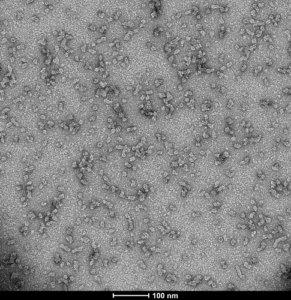
Figure 1. [Image from: StressMarq website]. Representative negative stain TEM image of StressMarq’s Kinetically-Stable Alpha Synuclein Oligomers (catalog# SPR-484).
Summary
Microglial activation is widely recognized as a key feature of Parkinson’s disease pathology, yet when it begins, how it relates to alpha synuclein aggregation, and the mechanisms that initiate it have remained poorly understood. Through a combination of transcriptomic analysis, single-cell profiling, and ultrasensitive mapping of alpha synuclein oligomers, Evans et al. have identified an early inflammatory phase of PD characterized by microglial activation and the accumulation of oligomeric alpha synuclein in cortical regions that lack detectable Lewy body pathology.
These findings suggest that immune activation and the presence of toxic alpha synuclein species may represent early, disease-driving events rather than secondary responses to neuronal degeneration. This work provides new insight into the molecular events that precede widespread pathology and highlights neuroinflammation and alpha synuclein oligomers as potential targets for early therapeutic intervention.
Related StressMarq products
StressMarq Biosciences manufactures a diverse portfolio of alpha synuclein, tau, amyloid beta, and TDP-43 proteins designed to support advanced preclinical neuroscience research. These include an innovative range of oligomeric, fibrillar and monomeric protein preparations, alongside antibodies, kits, and small molecules. Visit our website for more information, including the latest scientific publications using our specialized protein constructs.
References
- Microglial activation and alpha-synuclein oligomers drive the early inflammatory phase of Parkinson’s disease. Evans, J.R. et al. bioRxiv [Preprint]. 2025.
Leave a Reply