Deep Learning Models Transform PD Histopathology
Developing effective therapies for devastating neurodegenerative disorders such as Parkinson’s disease (PD) and Multiple System Atrophy (MSA) requires a thorough understanding of the cellular and molecular mechanisms that drive disease progression. In synucleinopathy research, preclinical studies rely heavily on histological analyses to characterize pathological changes and evaluate the efficacy of experimental interventions. Immunohistochemistry is widely used to assess protein aggregation, neuronal loss, and neuroinflammatory markers. However, these analyses are typically performed manually, making them labor-intensive, time-consuming, and susceptible to user bias and variability.
Recent advances in artificial intelligence (AI) are helping to address these limitations by enabling the automated, high-throughput analysis of histopathological samples. Researchers at KU Leuven in Belgium have successfully developed deep learning models capable of rapidly screening brain tissue for disease-specific pathological features. In a study published in NPJ Parkinson’s Disease, Barber-Janer and colleagues trained five convolutional neural network (CNN)-based models using mouse models of PD and MSA. Their platform employs a series of sequential CNNs, with each layer dedicated to executing a specialized task. The first network identifies brain tissue, the second maps specific brain regions, and subsequent networks are trained to detect individual pathological markers of interest.
Characterizing synucleinopaties
Both PD and MSA are characterized by the accumulation of misfolded alpha synuclein, although the cellular localization of these aggregates differs substantially between the two disorders. In PD, phosphorylated alpha synuclein (pSer129-aSyn) accumulates within neurons, forming abnormal intracellular deposits known as Lewy bodies (LBs) which are associated with neuronal dysfunction and degeneration in brain regions involved in movement, cognition, and memory. In contrast, MSA is characterized by the accumulation of alpha synuclein within oligodendrocytes, where it forms glial cytoplasmic inclusions (GCIs). These pathological aggregates contribute to widespread neurodegeneration affecting neural circuits responsible for motor control, balance, and autonomic function.
To generate disease-relevant animal models, the researchers delivered viral vectors encoding alpha synuclein to distinct cell populations. For the PD model, alpha synuclein expression was targeted to neurons within the substantia nigra (SN), whereas for the MSA model, expression was directed to oligodendrocytes in the dorsal striatum (DS). These complementary models enabled the authors to train and validate their CNN-based platform on disease-specific pathological features, establishing a powerful framework for automated histopathological analysis in synucleinopathy research.
Examining dopaminergic cells
A hallmark feature of Parkinson’s disease is the progressive loss of dopaminergic neurons within the SN. As a result, the quantification of dopaminergic neuron degeneration is a key endpoint in preclinical PD research and is commonly used to evaluate disease progression and therapeutic efficacy. In order to determine whether their AI-based platform could accurately detect neuronal loss, Barber-Janer et al. analyzed brain sections collected six months after viral vector injection. Tissue samples were immunohistochemically stained for tyrosine hydroxylase (TH), the rate-limiting enzyme in dopamine synthesis and a widely used marker of dopaminergic neurons.
Subsequently, the scientists employed a convolutional neural network trained to identify TH-positive cells within the substantia nigra. This dopaminergic cell detector (DCD) model was benchmarked against stereology, the current gold-standard method for quantifying neuronal populations. When applied to brain sections from both alpha synuclein-overexpressing and control mice, the DCD model generated results comparable to those obtained through stereological analysis while demonstrating greater consistency. Importantly, both approaches successfully distinguished between experimental groups, revealing a significant reduction in dopaminergic neuron numbers in alpha synuclein-overexpressing mice relative to controls.
Evaluating axonal degeneration
In addition to neuronal degeneration, axonal pathology is a prominent feature of PD and often precedes the loss of neuronal cell bodies. Degeneration of dopaminergic projections within the striatum is therefore considered a highly sensitive marker of early disease progression. To evaluate whether their platform could detect these more subtle pathological changes, the authors developed a second CNN-based model known as the dopaminergic axon detector (DAD). Rather than counting individual neurons, the DAD model quantified TH staining intensity within the dorsal striatum as a surrogate measure of dopaminergic axon density.
Using the same cohort of alpha synuclein-overexpressing and control mice, both the DAD model and conventional manual quantification methods detected a significant reduction in TH-positive axonal density in the experimental animals. Moreover, the strong correlation between the two approaches confirmed the accuracy and reliability of the CNN-based analysis. The ability to detect subtle alterations in axonal integrity is particularly valuable for preclinical research, as it enables the identification of pathological changes that emerge before overt neuronal loss becomes apparent. Together, the DCD and DAD models demonstrate that deep learning-based approaches can provide robust, high-throughput alternatives to traditional histological analyses, accelerating pathological assessment while maintaining the accuracy required for rigorous Parkinson’s disease research.
The role of neurons & microglia
Importantly, neurodegeneration in synucleinopathies extends beyond the substantia nigra and across multiple brain regions. To capture these broader pathological changes, the authors trained a neuronal cell detector (NCD) capable of identifying 28 distinct brain regions and quantifying neuronal density using NeuN immunostaining, a widely used marker of mature neurons.
The NCD was validated in the MSA mouse model, where alpha synuclein was overexpressed in oligodendrocytes. When compared with conventional stereological analysis, the model detected comparable levels of neuronal loss in the dorsal striatum and showed a strong correlation with manual measurements. Vitally, the NCD enabled large-scale brain-wide analysis that would be impractical using traditional methods. This approach revealed approximately 30% neuronal loss in the globus pallidus, 9% loss in the dorsal striatum, and additional subtle changes across multiple brain regions.
In addition to neurodegeneration, neuroinflammation is a prominent feature of both PD and MSA. To assess this aspect of synucleinopathy, the authors developed a microglial cell detector (MCD) trained to identify Iba1-positive microglia and evaluate their morphology. Using the same MSA mouse cohort, the MCD detected a 10 to 20% increase in microglial density across multiple brain regions, with region-specific patterns consistent with known alpha synuclein pathology. Similar to the NCD, the MCD enabled comprehensive brain-wide profiling of neuroinflammation that would otherwise require extensive manual analysis.
Appraising pSer129 pathology using StressMarq’s neurodegenerative disease constructs
Phosphorylated alpha synuclein is a key pathological hallmark of both PD and MSA, where it accumulates in fibrillar inclusions capable of recruiting endogenous alpha synuclein and propagating aggregation throughout the brain. To evaluate their pSer129-aSyn detector (pSynD), Barber-Janer et al. utilized StressMarq’s Mouse Alpha Synuclein Pre-formed Fibrils (catalog# SPR-324), a well-characterized fibrillar construct that has been shown to induce endogenous alpha synuclein aggregation following intracerebral injection. PFFs were injected into the dorsal striatum of mice, while control animals received PBS injections. The pSynD model was trained to identify the density, morphology, and anatomical distribution of pSer129-aSyn-positive inclusions in brain tissue.
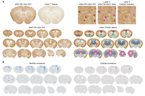
Figure 1. Unannotated representative pSer129-αSyn IHC and pSynD detection per layer: “Tissue” (layer 1, gray), “Brain region” (layer 2, different color for each brain region), “pSer129-αSyn+ area” (layer 3, blue for neuritic inclusions and magenta for cellular inclusions) and “Cellular inclusion” (layer 4, pink) and distribution of pSer129-αSyn+ neuritic inclusions (left, blue) and cellular inclusions (right, magenta) in a representative PFF-injected mouse. Figure taken from Barber-Janer et al., 2025 used under license CC BY 4.0.
Eight months after PFF injection, the pSynD detected a significant increase in neuritic pathology in PFF-treated mice compared with controls. In addition to quantifying pathology, the model mapped the brain-wide spread of alpha synuclein aggregates and identified regions with the highest densities of mature inclusions across 33 brain regions. The most heavily affected areas included the motor cortex, striatum, cingulate cortex, and amygdala. These findings demonstrate the utility of the pSynD model for high-throughput quantification of alpha synuclein pathology, providing a powerful tool for studying inclusion maturation, anatomical spread, and disease progression in preclinical synucleinopathy models.
Summary
Overall, Barber-Janer et al. have developed a powerful suite of CNN-based tools capable of automating brain-wide histopathological analyses in mouse models of synucleinopathy. Across multiple pathological endpoints, including neuronal loss, axonal degeneration, neuroinflammation, and alpha synuclein aggregation, the models closely matched the performance of conventional manual methods while offering substantially higher throughput, greater consistency, and reduced user-dependent variability. Tasks that would traditionally require weeks of manual analysis can now be completed in a fraction of the time.
Although these deep learning models were developed specifically for Parkinson’s disease and Multiple System Atrophy research, their utility extends beyond synucleinopathies. The ability to rapidly and accurately quantify pathological changes across entire brain sections makes this approach broadly applicable to a wide range of neurodegenerative and neuroinflammatory disease models. As artificial intelligence continues to be integrated into preclinical research workflows, tools such as these have the potential to accelerate discovery, improve reproducibility, and enhance the evaluation of emerging therapeutic strategies.
Related StressMarq Products
StressMarq Biosciences manufactures a diverse portfolio of alpha synuclein, tau, amyloid beta, and TDP-43 proteins designed to support advanced preclinical neuroscience research. These include an innovative range of oligomeric, fibrillar and monomeric protein preparations, alongside antibodies, kits, and small molecules. Visit our website for more information, including the latest scientific publications using our specialized protein constructs.
References
- Development of convolutional neural networks for automated brain-wide histopathological analysis in mouse models of synucleinopathies. Barber-Janer, A. et al. NPJ Parkinson’s Dis. 2025.
Leave a Reply