Advanced iPSC Systems to Model Alzheimer’s Disease Mechanisms
Alzheimer’s disease (AD) is the leading cause of dementia in older adults and remains a major public health challenge as populations age. Microglia are the brain’s resident innate immune cells, and are central to AD pathogenesis through roles in inflammation, synaptic remodeling, and phagocytosis of amyloid beta. Genetic mutations such as APOE, TREM2 and GRN alter microglial function and modulate disease risk, but how specific human genotypes change microglial responses to amyloid beta remains incompletely mapped. Translating animal models into effective human therapies has proved problematic, highlighting that preclinical research can fail to replicate human cell biology, particularly the contributions of microglia and human genetic risk variants.
In a research collaboration between Bioneer A/S, Denmark and StressMarq Biosciences, Canada, scientists utilized recent advances in induced pluripotent stem cell (iPSC) technology to develop human cellular models. These advanced models provide a more directly relevant platform for drug screening and studying the mechanisms of microglia‑mediated pathology.
Differentiation and validation of cell lines
Presented in a poster entitled: “Advanced Alzheimer’s Disease Models Using iPSC‑derived Microglia and Neurons Harboring Disease‑related Gene Variants”, Shatri et al.,(1) differentiated human iPSCs into microglia using CRISP/CAS-9 technology to introduce dementia-associated mutations. Alongside a wild-type cell line, the authors generated seven different mutant cell lines: APOE2/2, APOE3/3, APOE4/4, APOE knockout, TREM2R47H, GRN+/‑ and GRN knockout.
The APOE gene (apolipoprotein E) has three alleles, 2,3, and 4, each conveying different Alzheimer’s risk factors. APOE4/4 confers the highest Alzheimer’s risk, while APOE2/2 is thought to be neuroprotective.(2) Additionally, carriers of a specific TREM2R47H mutation in the TREM2 gene (triggering receptor expressed on myeloid cells 2) have an elevated risk of developing Alzheimer’s disease.(3) Loss-of-function mutations in GRN (progranulin) are primarily associated with frontotemporal lobar degeneration (FTLD) but may also play a role in AD.(4)
After generation, the mutant and wild-type cell lines were validated by testing for the presence of classic microglial markers. Immunostaining showed consistent expression of IBA‑1, TMEM119, TREM2 and P2Y12R across the edited lines, which confirmed a preserved microglial identity. The expression levels of the mutant genes in edited cell lines were also quantified. This validation established the model as both genetically faithful and phenotypically microglial, a prerequisite for downstream functional assays.
Immune response to cytokines and amyloid beta
The authors measured cytokine release by the different cell lines following stimulation with pathological triggers: StressMarq’s Amyloid Beta 1-42 Pre-formed Fibrils (catalog# SPR-487) and Amyloid Beta 1-42 Oligomers (catalog# SPR-488), or immune activators: IFN‑γ, LPS and TNF‑α.
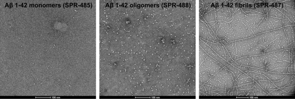
Figure 1. [Image from: StressMarq website] Transmission electron microscopy (TEM) of Amyloid Beta 1-42 Monomers (catalog# SPR-485, left), Amyloid Beta 1-42 Oligomers (catalog# SPR-488, middle) and Amyloid Beta 1-42 Pre-formed Fibrils (catalog# SPR-487, right).
Crucially, TREM2R47H microglia did not mount the same cytokine response to amyloid beta oligomers, indicating that this AD‑linked TREM2 variant either dampens or alters microglial sensing or signaling pathways in response to oligomeric amyloid beta. Researchers also measured dose responsiveness in response to graded doses of IFN‑γ, TNF‑α and LPS.
Phagocytosis of amyloid beta and E. coli
Phagocytic competence is a key microglial function relevant to amyloid beta clearance. Using pHrodo‑conjugated amyloid beta oligomers and E. coli particles, the authors monitored real‑time phagocytic uptake across APOE genotypes. pHrodo is a dye that becomes more fluorescent in acidic environments, such as inside phagosomes. This property makes it useful for tracking phagocytosis.
APOE3/3 microglia showed higher uptake of amyloid beta oligomers compared with APOE4/4 and APOE KO, suggesting that the APOE genotype influences microglial capacity to internalize oligomeric amyloid beta. Given the established association of APOE4 with greater AD risk, reduced phagocytic removal of oligomers by APOE4 microglia could be a functional contributor. However, all lines retained the basic ability to phagocytose bacterial particles and amyloid beta despite genetic differences, highlighting how genotype shifts efficiency rather than abolishing function.
To increase physiological relevance, the group adapted existing co‑culture protocols combining NGN2‑induced neurons with their iPSC‑derived microglia. Immunostaining confirmed the presence of neuronal markers (β‑Tubulin, MAP2) alongside microglial markers (IBA‑1, TREM2, TMEM119) in co‑cultures – enabling examination of microglial responses in the presence of neuronal signals and monitoring of cellular interactions.
Summary
Shatri et al. present validated, practical protocols for producing iPSC‑derived microglia and NGN2‑induced neuron co‑cultures carrying Alzheimer’s‑related gene edits. Their microglial models exhibit expected marker profiles and functional behaviors, and display genotype‑specific alterations in cytokine release and phagocytic activity. This human‑relevant, genotype‑aware co‑culture offers a robust platform for drug discovery and neurodegeneration research, enabling screening of compounds that modulate microglial inflammation and promote amyloid‑beta clearance.
Related StressMarq products
StressMarq’s range of high-quality protein preparations and antibodies for amyloid beta, tau, and alpha synuclein provide a complete toolkit for the study of amyloidogenic protein misfolding and neurotoxicity. The Amyloid Beta Pyroglutamate 3-42 Pre-formed Fibrils (catalog# SPR-492) deliver disease‑relevant, highly stable fibrils, while the Anti-Amyloid Beta Oligomer antibodies (catalog# SMC-618, SMC-619 and SMC-620) enable sensitive detection of toxic oligomeric amyloid beta species in a variety of immunoassays.
References
- Advanced Alzheimer’s Disease Models Using iPSC-derived Microglia and Neurons Harboring Disease-related Gene Variants. Shatri, E.B. et al. (Poster).
- ApoE in Alzheimer’s disease: pathophysiology and therapeutic strategies. Raulin, AC. et al. Mol Neurodegener. 2022.
- Variant of TREM2 associated with the risk of Alzheimer’s disease. Jonsson, T. et al. N Engl J Med. 2013.
- Progranulin haploinsufficiency reduces amyloid beta deposition in Alzheimer’s disease model mice. Hosokawa, M. et al. Exp Anim. 2018.
Leave a Reply