
Tau Pathology and the Circadian Pacemaker
Tauopathies are a group of neurodegenerative disorders defined by the abnormal folding, hyperphosphorylation, and aggregation of the microtubule‑associated protein tau. Conditions in this family, such as Alzheimer’s disease (AD) cause progressive cognitive decline, synaptic failure, and neuronal loss. Alongside memory and behavioral symptoms, patients frequently develop severe circadian disturbances as the disease advances.
The suprachiasmatic nucleus (SCN) in the hypothalamus is the brain’s master circadian pacemaker. It takes external environment cues, or Zeitgebers, and synchronizes the circadian clock with the outside world. This process is known as entrainment. Light is the dominant Zeitgeber, thus it is the daily light-dark cycle that most strongly influences circadian rhythms. Patients in the later stages of neurodegeneration suffer from fragmented sleep, irregular activity-rest patterns, and progressive day-night disorganization. Not only do these changes degrade their quality of life, but they can potentially accelerate neurodegeneration. Post‑mortem studies of AD patients have reported tau pathology in the human SCN, but whether tau accumulation directly disrupts the SCN’s timing machinery has remained unclear.
To better understand tau in the SCN, researchers from The Ohio State University, USA, employed the transgenic PS19 mouse model to study the effect of tau accumulation. This murine AD model overexpresses human tau carrying the P301S mutation and recapitulates key features of human tauopathies, including tau hyperphosphorylation, aggregation, neurofibrillary tangle (NFT) formation, and neuronal loss. Importantly, it exhibits a well-characterized, age-dependent progression of tau pathology, with early gliosis and synaptic loss evident by 3 months, NFTs developing by 6 months, and substantial neuronal loss by 9 months.
Early accumulation of pathogenic tau
Published in Experimental Neurology, Halloy et al. began by investigating transgenic tau expression. Using the well-characterized AT8 tau antibody, which recognizes tau phosphorylated at Serine 202 and Threonine 205, the group mapped phosphorylated tau (p‑tau) across ages and brain regions. As expected, cortical areas showed progressive accumulation with age. Crucially, the SCN displayed diffuse but marked p‑tau labeling as early as 2 months of age, a time point that precedes many overt pathological changes elsewhere in the central nervous system. A subset of SCN neurons developed intense AT8 staining consistent with tau‑positive inclusions, although overall labeling intensity showed a complex pattern across ages.
In addition to the SCN, the authors quantified p‑tau in the piriform cortex, striatum, and retrosplenial cortex. Each region followed a distinct temporal profile. The piriform cortex showed low early expression that escalated by 11 months, while the striatum and retrosplenial cortex exhibited minimal early pathology but marked accumulation over time. These region‑specific differences demonstrate that tauopathy progresses unevenly across the brain and that the SCN is an unusually early site of tau deposition in this model. Fluorescent co‑labeling revealed that p‑tau was present in both major SCN neuronal subpopulations – the core and the shell. This indicates that tau pathology is not restricted to a single SCN compartment but that it affects the principal cell types that coordinate pacemaker function.
Role of clock-timing and entrainment in the SCN
The SCN pacemaker works by molecular oscillation, coordinating peripheral circadian oscillator populations across all organs. It generates these rhythmic outputs by transcriptional-translational feedback loops of core clock genes, which include Clock, Bmal1, Per1/2 and Cry1/2. To test whether SCN tau pathology translated into altered circadian behavior, Halloy et al. monitored locomotor wheel running activity in PS19 and wild‑type (WT) mice from 3 to 11 months of age.
The researchers assessed multiple aspects of circadian function, including activity under standard light/dark cycles (Fig. 1), ability to adapt (re‑entrain) to 6‑hour phase advances and delays, and intrinsic pacemaker properties under constant darkness (DD) and constant light (LL). In all these settings, PS19 mice were indistinguishable from controls. Even under the stress of constant light, which can challenge circadian stability, PS19 animals maintained robust rhythms. This indicates that SCN‑driven locomotor timing is remarkably resilient to tau accumulation in this model.
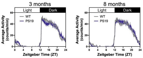
Figure 1: [From Halloy et al. 2025.] Circadian-gated locomotor activity in PS19 mice. Mean daily activity profiles of WT and PS19 mice kept in phased 12-hour light/dark cycles at 3 months and 8 months old.
Effect of tau on clock gene expression
To further investigate the effect of pathogenic tau on the SCN, the group cultured SCN explants from PS19 and WT mice expressing the Per1-Venus circadian reporter. In these transgenic lines, the Per1 clock gene drives expression of a fluorescent Venus reporter, enabling real-time visualization of circadian gene activity. The researchers isolated the SCN tissue from PS19 and WT mice, and maintained the tissue under confocal microscopy, imaging over several weeks to track Per1‑driven fluorescence rhythms. Interestingly, this revealed that circadian oscillations in Per1 expression were comparable between PS19 and WT cultured explants.
To accelerate tau pathology, both WT and PS19 explants were seeded with StressMarq’s Tau-441 (2N4R) P301S Mutant Pre-formed Fibrils (catalog# SPR-329) at two different concentrations. The researchers confirmed tau accumulation by immunolabelling with AT8 and MC1 antibodies against p-tau and paired helical filaments, respectively. Analysis of circadian period, amplitude, and rhythmicity revealed no consistent differences between PS19 and wild‑type explants. Additionally, tau seeding did not produce measurable disruptions in oscillator properties. These findings reinforced the behavioral data. Taken together, these findings indicated that clock timing and network synchrony in this SCN model are resilient to the presence of substantial tau pathology.
Summary
Using their PS19 transgenic mouse model, Halloy et al. demonstrated that despite the early and progressive deposition of tau in the SCN, core circadian timing functions were not impaired. Transgenic PS19 mice maintained normal circadian locomotor rhythms in vivo. In addition, ex vivo SCN explants preserved robust oscillator properties, even after successful tau seeding. These findings challenge the assumption that circadian dysfunction in tauopathies stems directly from SCN impairment. Instead, the authors propose that disruptions may arise from SCN output pathways or downstream clock-gated processes.
Related StressMarq products
StressMarq Biosciences offers researchers a comprehensive portfolio of high-quality tau, alpha synuclein, amyloid beta, and TDP-43 proteins. Designed to deliver exceptional performance in neurodegenerative disease research, this diverse catalog of cutting-edge reagents undergoes rigorous validation to ensure purity, consistency, and biological relevance, enabling scientists to model proteinopathies with confidence and generate reproducible, publication-ready data. Visit our website for more information, including the latest scientific publications using our specialized oligomeric, fibrillar and monomeric protein preparations, as well as our antibodies, kits, and small molecules.
References
- Circadian timing and entrainment properties of the SCN pacemaker in the PS19 mouse model of tau pathology. Halloy, N. R. et al. Exp Neurol. 2026.
Leave a Reply