Advancing the Frontiers of Neurodegenerative Disease Research


Properties
| Storage Buffer | PBS pH7.4, 50% glycerol, 0.09% sodium azide *Storage buffer may change when conjugated |
| Storage Temperature | -20ºC, Conjugated antibodies should be stored according to the product label |
| Shipping Temperature | Blue Ice or 4ºC |
| Purification | Protein G Purified |
| Clonality | Monoclonal |
| Clone Number | L60/4 (Formerly sold as S60-4) |
| Isotype | IgG2b |
| Specificity | Detects ~180kDa in rat brain membrane preparations. |
| Cite This Product | ATP7A Antibody (StressMarq Biosciences | Victoria, BC CANADA, Catalog# SMC-398, RRID: AB_11232613) |
| Certificate of Analysis | 1 µg/ml of SMC-398 was sufficient for detection of Copper-transporting ATPase1 in 20 µg of rat brain lysate by colorimetric immunoblot analysis using Goat IgG:HRP as the secondary antibody. |
Biological Description
| Alternative Names | ATP7A, ATP 7A, ATP7A_HUMAN, ATPase Cu transporting, ATPase copper transporting alpha polypeptide, ATPase Cu++ transporting alpha polypeptide (Menkes syndrome), Copper transporting ATPase 1, Cu++ transporting P type ATPase, Menkes disease associated protein, Menkes syndrome, Menke, MC1, MC 1, MK, MNK, DSMAX, SMAX3, OHS, FLJ17790, OTTHUMP00000062077 |
| Research Areas | Cell Signaling, Neurodegeneration, Neuroscience |
| Cellular Localization | Cell membrane, Cytoplasm, Endoplasmic Reticulum, Golgi apparatus, Trans-golgi network membrane |
| Accession Number | NP_000043.3 |
| Gene ID | 538 |
| Swiss Prot | Q04656 |
| Scientific Background |
ATP7A, also known as copper-transporting ATPase 1, is a transmembrane P-type ATPase that plays a critical role in cellular copper homeostasis. It facilitates the export of excess intracellular copper by trafficking it into the secretory pathway for vesicular exocytosis. This function is essential for systemic copper absorption in the intestine and reabsorption in the kidney, processes that are effectively modeled in polarized epithelial systems such as Madin-Darby canine kidney (MDCK) cells. In the nervous system, ATP7A is indispensable for delivering copper to cuproenzymes involved in neurotransmitter synthesis, antioxidant defense, and mitochondrial function. Disruption of ATP7A-mediated copper transport leads to severe neurodevelopmental consequences, as seen in Menkes disease, a fatal X-linked disorder characterized by copper deficiency, neurodegeneration, and connective tissue abnormalities. Emerging research links ATP7A dysfunction to broader neurodegenerative processes. Impaired copper regulation contributes to oxidative stress, protein misfolding, and mitochondrial dysfunction—hallmarks of diseases such as Alzheimer’s and Parkinson’s. Moreover, ATP7A expression is altered in various cancers and has been associated with resistance to platinum-based chemotherapies like cisplatin and carboplatin. Given its dual role in systemic copper regulation and neuronal health, ATP7A is a promising target for therapeutic strategies aimed at restoring metal homeostasis in neurodegenerative diseases. Understanding its regulation and trafficking dynamics may unlock new avenues for intervention in copper-related neuropathologies. |
| References |
1. Samimi G., et al. (2003) Clin. Cancer Res. 9: 5853-9. 2. Samimi G., et al. (2004) Mol Pharmacol. 66: 25-32. 3. Greenough M., et al. (2004) Am. J. Physiol. Cell Physiol. 287: C1463-71. 4. Song, I.S., et al. (2004) Mol. Cancer Ther. 3: 1543-1549. 5. van Dongen, E.M., et al. (2004) Biochem. Biophys. Res. Commun. 323: 789-795. 6. Samimi, G., et al. (2004) Mol Pharmacol 66: 25-32. 7. Morgan, C.T., et al. (2004) J. Biol. Chem. 279: 36363-36371. 8. Barnes, N., et al. (2005) J. Biol. Chem. [Epub]. |
Product Images
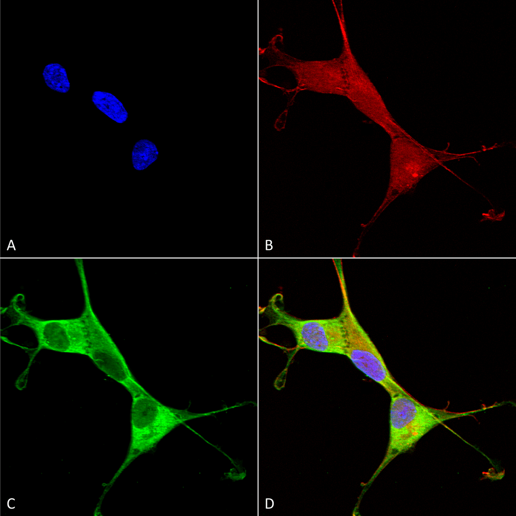
Immunocytochemistry/Immunofluorescence analysis using Mouse Anti-Copper Transporting ATPase 1 Monoclonal Antibody, Clone L60/4 (SMC-398). Tissue: Neuroblastoma cells (SH-SY5Y). Species: Human. Fixation: 4% PFA for 15 min. Primary Antibody: Mouse Anti-Copper Transporting ATPase 1 Monoclonal Antibody (SMC-398) at 1:100 for overnight at 4°C with slow rocking. Secondary Antibody: AlexaFluor 488 at 1:1000 for 1 hour at RT. Counterstain: Phalloidin-iFluor 647 (red) F-Actin stain; Hoechst (blue) nuclear stain at 1:800, 1.6mM for 20 min at RT. (A) Hoechst (blue) nuclear stain. (B) Phalloidin-iFluor 647 (red) F-Actin stain. (C) Copper Transporting ATPase 1 Antibody (D) Composite.
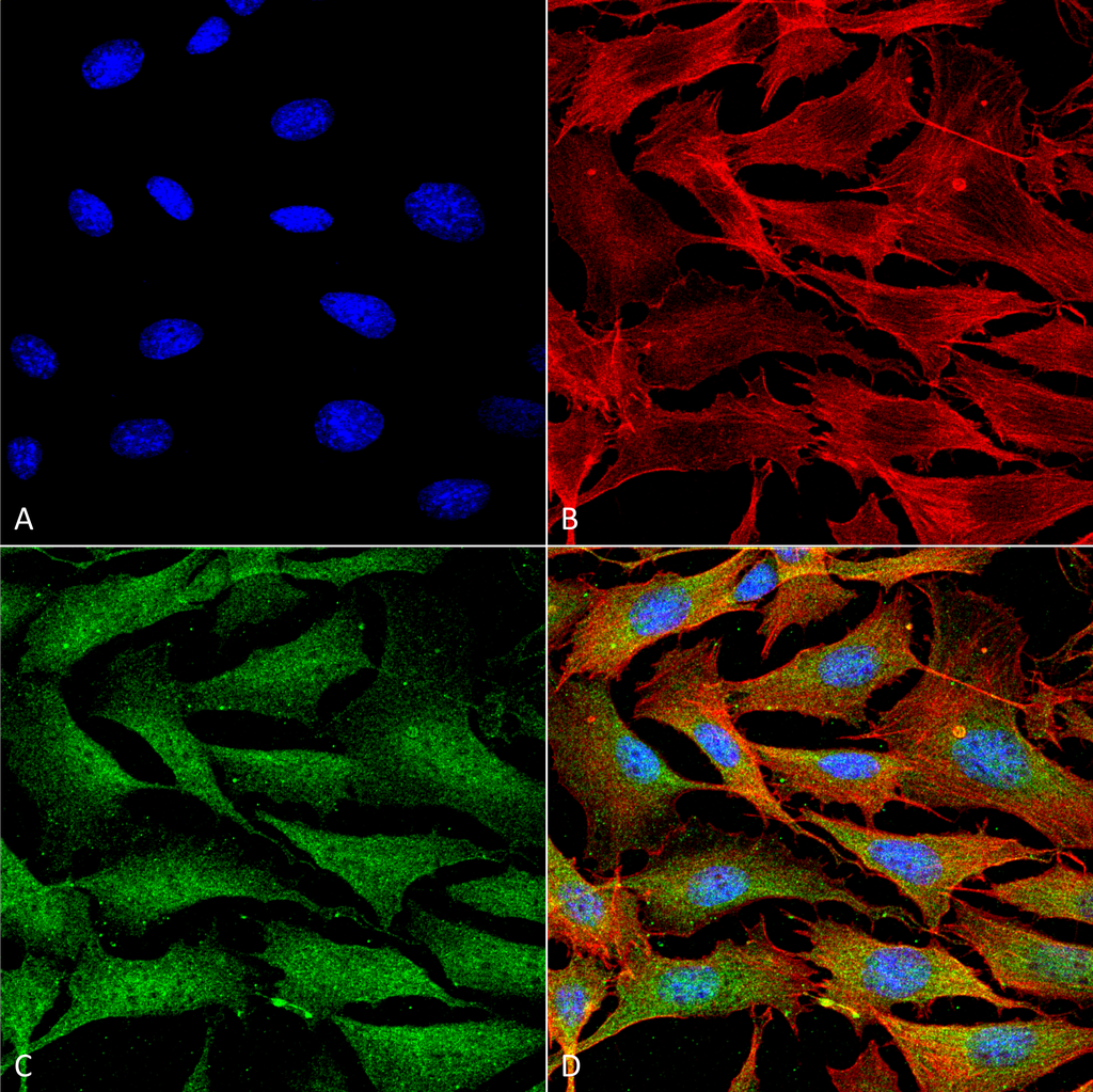
Immunocytochemistry/Immunofluorescence analysis using Mouse Anti-Copper Transporting ATPase 1 Monoclonal Antibody, Clone L60/4 (SMC-398). Tissue: NIH 3T3 (NIH 3T3). Species: Mouse. Fixation: 4% Formaldehyde for 15 min at RT. Primary Antibody: Mouse Anti-Copper Transporting ATPase 1 Monoclonal Antibody (SMC-398) at 1:100 for 60 min at RT. Secondary Antibody: Goat Anti-Mouse ATTO 488 at 1:200 for 60 min at RT. Counterstain: Phalloidin Texas Red F-Actin stain; DAPI (blue) nuclear stain at 1:1000, 1:5000 for 60 min at RT, 5 min at RT. Localization: Endoplasmic Reticulum, Cytoplasm, Golgi Apparatus, Trans-Golgi Network Membrane, Cell Membrane. Magnification: 60X. (A) DAPI (blue) nuclear stain. (B) Phalloidin Texas Red F-Actin stain. (C) Copper Transporting ATPase 1 Antibody. (D) Composite.
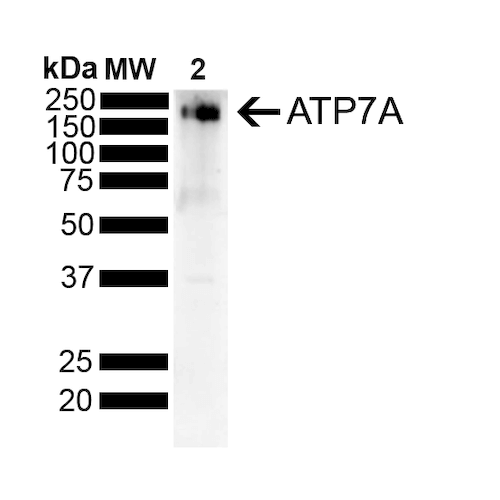
Western Blot analysis of Rat Liver showing detection of ~180 kDa Copper Transporting ATPase 1 protein using Mouse Anti-Copper Transporting ATPase 1 Monoclonal Antibody, Clone L60/4 (SMC-398). Lane 1: MW Ladder. Lane 2: Rat Liver . Load: 10 ug. Block: 5% Skim Milk powder in TBST. Primary Antibody: Mouse Anti-Copper Transporting ATPase 1 Monoclonal Antibody (SMC-398) at 1:500 for 2 hours at RT with shaking. Secondary Antibody: Goat anti-mouse IgG:HRP at 1:4000 for 1 hour at RT with shaking. Color Development: Chemiluminescent for HRP (Moss) for 5 min in RT. Predicted/Observed Size: ~180 kDa.
 Powered by Bioz
Reviews
There are no reviews yet.