
Alpha Synuclein and Tau Cross-Seeding in Neurodegenerative Disease
Neurodegenerative diseases such as Parkinson’s disease (PD), Alzheimer’s disease (AD), and dementia with Lewy bodies (DLB), are characterized by the abnormal accumulation of misfolded proteins in the brain, leading to progressive cognitive and motor decline. The pathological deposition of alpha synuclein protein aggregates is the hallmark of synucleinopathies such as Parkinson’s disease and DLB, whereas the deposition of tau is the defining feature of tauopathies including Alzheimer’s disease. Interestingly, these proteins have been observed to co-deposit in patient brains in both Alzheimer’s and Parkinson’s disease.
Post-mortem studies reveal that up to 50% of Alzheimer’s patients also show alpha synuclein deposits, and tau pathology is frequently observed in Parkinson’s disease. This overlap suggests a pathological link between disorders that have traditionally been considered distinct. A key mechanism thought to drive this overlap is cross-seeding — the ability of one misfolded protein to accelerate the aggregation of another.
Under pathological conditions the disordered, soluble monomeric forms of tau and alpha synuclein adopt extensive β-sheet structures. These structures lead to the formation of insoluble fibrils enriched with stacked β-sheets. Previous studies have shown structural diversity between these aggregates and corresponding differences in seeding ability. However, until now, scientists lacked the tools to directly visualize and characterize the structural diversity of these aggregates inside living cells.
In a collaboration between China Medical University, Lund University, Light Chain Bioscience, Discoveric Bio and StressMarq Biosciences, researchers utilized optical photothermal infrared (O‑PTIR) microspectroscopy combined with confocal imaging to study the mechanism of alpha synuclein and tau cross-seeding with different strain-specific seeds.
The tau isoform affects morphology of co-polymers
Tau exists in six major isoforms in the human brain, generated by alternative splicing of the MAPT gene. They differ in the number of N-terminal inserts (0N, 1N or 2N) and the number of microtubule-binding repeats (3R or 4R). Together, these combinations yield 0N3R, 1N3R, 2N3R, 0N4R, 1N4R, and 2N4R isoforms.
Published in the Journal of the American Chemical Society, Zhan et al. characterized several fibril preparations obtained from StressMarq Biosciences. In addition to Alpha Synuclein Monomers (catalog# SPR-321), the group looked at preparations of Alpha Synuclein Pre-formed Fibrils (catalog# SPR-322), tau 3R and 4R isoforms – Tau-352 (fetal 0N3R) Wild-Type Pre-formed Fibrils (catalog# SPR-491) and Tau-441 (2N4R) Wild-Type Pre-formed Fibrils (catalog# SPR-480). As well, they used alpha synuclein/tau3R Tau and Alpha Synuclein Co-Polymer Fibrils (catalog# SPR-494) and alpha synuclein/tau4R Tau and Alpha Synuclein Co-Polymer Fibrils (catalog# SPR-495).
Atomic force microscopy (AFM) revealed that co-polymers were thicker and more ribbon-like than pure fibrils. Additionally, the researchers employed immuno-TEM (immunogold transmission electron microscopy) utilizing StressMarq’s Anti-Alpha Synuclein Antibody (catalog# SMC-532) and Anti-Tau Antibody (catalog# SPC-801). This showed that alpha synuclein/tau3R contained nearly equal amounts of alpha synuclein and tau, while alpha synuclein/tau4R was heavily tau dominant. These findings suggested that the tau isoform strongly influences the structural balance of the resulting fibrils.
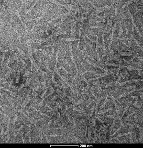
Figure 1: [Image from StressMarq website] Transmission electron microscopy (TEM) of Tau 2N4R & Alpha Synuclein Co-Polymer Fibrils (catalog# SPR-495).
While alpha synuclein/tau3R exhibited the highest β‑sheet content, tau4R-containing hybrids, by contrast, showed weaker β‑sheet enrichment and more random coil content. Through principal component analysis (PCA) the researchers confirmed that each fibril type had a distinct structural fingerprint. This demonstrated that tau isoform identity dictates the structural outcome of alpha synuclein/tau hybrids, with tau3R driving more amyloidogenic folds.
Seeding activity is related to template fibril composition
To test the seeding characteristics of each fibril type, the researchers seeded HEK293 cells expressing fluorescently tagged A53T mutant alpha synuclein with the 7 different pre-formed fibrils and alpha synuclein monomers. The scientists then monitored inclusion formation over 24 to 48 hours. During this period they visualized the inclusions by accumulation of fluorescent alpha synuclein within the cells. After 12 hours no inclusions were visible for cells treated with alpha synuclein monomers or tau pre-formed fibrils. However, after 48 hours, alpha synuclein/tau3R seeds had produced the most robust seeding activity, and a higher percentage of these cells developed inclusions which were larger and more intense. Cytotoxicity assays confirmed that these seeds were also the most toxic. Thus, structural differences detected by O‑PTIR translated directly into functional differences in cellular seeding potency and toxicity.
Confocal imaging of cells revealed that inclusions induced by different seeds varied in both morphology and post-translational modifications. Alpha synuclein/tau3R inclusions showed the highest phosphorylation at p‑S129 (alpha synuclein) and AT8 (tau) as detected by immunofluorescence. Colocalization analysis confirmed a strong overlap between phosphorylated alpha synuclein and tau within inclusions seeded by tau3R hybrids. Notably, 3D reconstructions highlighted distinct spatial patterns of colocalization depending on seed type. Further, biochemical fractionation revealed additional differences in solubility and protease resistance.
In contrast, the alpha synuclein/tau4R inclusions were less resistant to proteinase K, reflecting weaker β‑sheet enrichment. Using confocal imaging and corrective O-PTIR, the researchers identified structural heterogeneity between the inclusions. Aggregates retained the structure of their seed templates, with alpha synuclein/tau3R seeded cells exhibiting the highest levels of β‑sheet structure. These results emphasize that cellular inclusions inherit structural motifs from their donor seeds, which leads to divergent properties.
Summary
By combining O‑PTIR spectroscopy with confocal imaging, Zhan et al. achieved sub-cellular resolution structural mapping of amyloid proteins directly in cells. Thus providing compelling evidence that alpha synuclein and tau cross-seeding generates structurally distinct and functionally divergent amyloid strains. The discovery that alpha synuclein/tau3R hybrids are particularly potent in driving β‑sheet enrichment, seeding, and phosphorylation highlights the importance of tau isoform composition in disease mechanisms. These findings suggest that strain diversity in alpha synuclein/tau aggregates could underlie the clinical variability seen across neurodegenerative disorders.
Related StressMarq products
StressMarq Biosciences offers a comprehensive portfolio of pre-formed fibrils for critical proteins in neurodegeneration research, such as tau, alpha synuclein, and amyloid beta. These rigorously validated reagents provide researchers with reliable tools to model protein aggregation, cross‑seeding, and strain diversity. With tau isoforms (3R and 4R) to study tauopathies, alpha synuclein fibrils to investigate synucleinopathies, and amyloid beta fibrils to explore Alzheimer’s pathology, StressMarq’s products enable scientists to examine the molecular mechanisms underlying neurodegenerative disease and advance translational research.
References
- Strain-Distinct α-Synuclein and Tau Cross-Seeding Uncovered by Correlative Approach with Optical Photothermal Infrared Sub-Micron Imaging. Zhan, X. et al. J Am Chem Soc. 2025;147(31):27323-27340.
Leave a Reply